
Overview
Acute deep vein thrombosis is a blood clot that forms in a deep vein, most often in the leg or pelvis. It is one of the two faces of venous thromboembolism (VTE), the other being pulmonary embolism (PE), when part of the clot travels to the lungs. DVT matters for two reasons. In the short term it can cause a life-threatening PE. In the long term it can damage the vein and cause chronic leg swelling, pain and skin changes (post-thrombotic syndrome).
The clot forms through the three factors of Virchow’s triad: venous stasis (immobility, venous obstruction), endothelial injury (surgery, trauma, catheters) and hypercoagulability (cancer, pregnancy, oestrogens, thrombophilia). Most patients have more than one of these.
DVT is classified by location:
- Distal (calf) DVT: the calf veins below the knee. It has a lower risk of embolisation but can extend.
- Proximal DVT: the popliteal, femoral, common femoral and iliac veins. This carries most of the risk of PE and post-thrombotic syndrome.
- Iliofemoral DVT: thrombus in the iliac and common femoral veins. It is the most symptomatic and has the highest risk of long-term venous damage. It is more common on the left, partly because the right common iliac artery crosses over the left common iliac vein (May-Thurner anatomy).
- Upper limb DVT: about 5–10% of cases. It is usually catheter-related or associated with cancer, or is effort thrombosis (Paget-Schroetter syndrome), often on a background of thoracic outlet compression.
How common is it?
A prospective community study in north-eastern Perth found a crude annual incidence of 0.52 per 1000 residents for DVT and 0.83 per 1000 for VTE overall, which extrapolates to roughly 17,000 Australians with VTE each year. International studies, including Australian data, report annual VTE incidences of 0.75 to 2.69 per 1000, rising to 2–7 per 1000 in people aged 70 or over. The Thrombosis and Haemostasis Society of Australia and New Zealand cites a lifetime VTE risk of about 8%, and it is a recurrent condition for many people.
Incidence rises steeply with age. It is also strongly linked to hospitalisation, and much hospital-associated VTE is preventable with risk assessment and appropriate prophylaxis. Australian hospital data suggest VTE accounts for around 7% of in-hospital deaths.
Risk factors
| Category | Examples |
|---|---|
| Major transient (provoking) | Surgery (especially orthopaedic, cancer, pelvic), hospitalisation with acute illness, major trauma or lower limb fracture, prolonged immobility or bed rest, Caesarean section |
| Minor transient | Oestrogen-containing contraception or HRT, pregnancy and the puerperium, long-haul travel (a modest risk increase), central venous catheters, minor leg injury, dehydration |
| Persistent | Active cancer and chemotherapy, previous VTE, obesity, older age, inflammatory bowel disease, nephrotic syndrome, heart failure, myeloproliferative neoplasms |
| Thrombophilia | Factor V Leiden and prothrombin gene mutation (modest risk); protein C, protein S or antithrombin deficiency (higher risk); antiphospholipid syndrome (important for treatment choice) |
| Anatomical | May-Thurner (iliac vein compression), other venous compression syndromes, previous venous damage |
About half of first DVTs occur with an identifiable provoking factor. The remainder are classed as “unprovoked”, which has real consequences for how long treatment continues.
Clinical presentation
Symptoms and signs
- Unilateral leg swelling, the most consistent finding
- Pain or tenderness, often calf or thigh, described as aching, tightness or cramping
- Warmth, redness or bluish discolouration
- Pitting oedema confined to one leg
- Distended superficial collateral veins
- Calf circumference more than 3 cm greater than the other side, measured 10 cm below the tibial tuberosity
DVT can also be silent. Many patients with proximal DVT have no leg symptoms at all, and concurrent asymptomatic PE is common. Homan’s sign is unreliable and should not be used.
Severe presentations (urgent)
- Phlegmasia alba dolens: a pale, painful, swollen leg from massive iliofemoral thrombosis.
- Phlegmasia cerulea dolens: a tense, blue, very painful leg with impending venous gangrene. It can compromise arterial inflow and is a limb- and life-threatening emergency.
Features suggesting PE: sudden shortness of breath, pleuritic chest pain, haemoptysis, palpitations, presyncope or syncope. These need emergency assessment.
Differential diagnosis: cellulitis, ruptured Baker’s cyst, muscle strain or haematoma, chronic venous insufficiency, lymphoedema or lipoedema, heart or renal failure (usually bilateral), superficial thrombophlebitis, compartment syndrome and acute arterial ischaemia.
Investigations
1. Estimate pre-test probability (Wells score). One point each for:
- active cancer
- paralysis or recent plaster immobilisation
- bedridden more than 3 days or major surgery within 12 weeks
- localised tenderness along the deep venous system
- entire leg swollen
- calf swelling more than 3 cm
- pitting oedema
- non-varicose collateral superficial veins
- previous documented DVT
Subtract 2 if an alternative diagnosis is at least as likely. A score of 2 or more means “DVT likely”; below 2 means “DVT unlikely”.
2. D-dimer (if DVT unlikely). A negative high-sensitivity D-dimer effectively excludes DVT. A positive result is non-specific and needs imaging. It is also raised in pregnancy, cancer, infection, recent surgery and older age, so it is often unhelpful in those groups.
3. Compression duplex ultrasound (if DVT likely, or D-dimer positive). This is the first-line imaging test. The key finding is a vein that will not compress under the probe. Supporting findings are echogenic intraluminal thrombus, loss of normal flow and respiratory variation, and venous distension. Sensitivity for proximal DVT exceeds 95%. It is lower for calf veins, and either a whole-leg scan or a repeat proximal scan at 5–7 days is used when suspicion persists.
4. Imaging of the iliac veins and IVC. Ultrasound has limits in the pelvis and abdomen. When iliofemoral or caval thrombosis is suspected (for example, absent respiratory phasicity in the common femoral vein, or thigh-to-groin swelling with a negative leg scan), CT or MR venography is used. Venography with intravascular ultrasound (IVUS) is used in the interventional setting.
5. Baseline blood tests. FBC, renal function, liver function and coagulation screen before anticoagulation, as they guide drug choice and dosing.
6. Thrombophilia and cancer assessment. Broad thrombophilia testing is not routine. It is reserved for selected patients, such as young people with unprovoked events, a strong family history, unusual sites, or recurrence. Testing during acute thrombosis or on anticoagulants can give misleading results. Testing for antiphospholipid syndrome matters because DOACs are less effective in high-risk (triple-positive) disease, where warfarin is preferred. Occult cancer assessment should be limited to history, examination, routine bloods and age-appropriate screening. Extensive whole-body imaging has not been shown to improve outcomes.
Management
Anticoagulation: the foundation of treatment
Treatment aims to stop clot extension, prevent PE, reduce recurrence and let the body remodel the clot. Almost all patients with acute proximal DVT need therapeutic anticoagulation.
- Direct oral anticoagulants (DOACs) are now first-line for most patients (CHEST 2021, ESVS 2021, ASH 2020). Apixaban is given at 10 mg twice daily for 7 days, then 5 mg twice daily. Rivaroxaban is given at 15 mg twice daily for 21 days, then 20 mg daily with food. Dabigatran and edoxaban require an initial 5–10 days of parenteral anticoagulation.
- LMWH bridged to warfarin (INR 2–3) remains appropriate in some situations, including antiphospholipid syndrome, severe renal impairment and some drug interactions.
- Unfractionated heparin is used when rapid reversibility is needed, in severe renal impairment, or when intervention is being considered.
- Cancer-associated DVT: a DOAC (apixaban, edoxaban or rivaroxaban) or LMWH, with attention to GI and genitourinary tumours because of bleeding risk.
- Pregnancy: LMWH throughout pregnancy and for at least 6 weeks postpartum. DOACs and warfarin are avoided.
Most uncomplicated DVT can be managed at home. Admission is considered for severe symptoms, phlegmasia, suspected haemodynamically significant PE, high bleeding risk, renal failure or limited social support.
Duration of anticoagulation
| Situation | Suggested approach |
|---|---|
| DVT provoked by a major transient factor (for example, surgery) | 3 months, then stop |
| DVT with a minor transient factor | 3 months, then individual review (a longer course may be reasonable) |
| Unprovoked DVT or persistent risk factor | Extended (indefinite) therapy considered if bleeding risk is acceptable, reviewed at least annually. After 6 months, reduced-dose apixaban (2.5 mg twice daily) or rivaroxaban (10 mg daily) is an option |
| Active cancer | Continue while the cancer is active or under treatment |
| Isolated distal DVT | 3 months if symptomatic or at risk of extension. Serial ultrasound surveillance is an option for mild symptoms and low risk |
Compression and mobilisation
Early walking is safe and encouraged. Graduated compression stockings can relieve pain and swelling once the acute pain settles. However, in the SOX trial, routine stockings did not prevent post-thrombotic syndrome, so they are used for symptoms rather than as routine prevention.
Advanced (interventional) therapy
Anticoagulation does not actively remove clot. Removing it early may protect the vein and its valves. This is a vascular surgical decision, considered in selected patients with:
- iliofemoral (or extensive proximal) DVT with severe symptoms
- symptom onset within roughly 14 days
- low bleeding risk and reasonable functional status
- or phlegmasia cerulea dolens or a threatened limb, where intervention is urgent
Options include:
- Catheter-directed thrombolysis (CDT), delivering clot-dissolving drug directly into the thrombus
- Pharmacomechanical thrombolysis, combining lytic drug with mechanical disruption or ultrasound acceleration
- Percutaneous mechanical thrombectomy, removing thrombus with aspiration or rheolytic devices, often without lytic drugs
- Balloon angioplasty and venous stenting, treating an underlying stenosis such as May-Thurner syndrome once the clot has been cleared
- Open surgical venous thrombectomy, rarely, for limb-threatening cases
The evidence is mixed and needs to be discussed candidly. In CaVenT, CDT reduced post-thrombotic syndrome at two years (about 41% versus 56%). The larger ATTRACT trial found no difference in overall post-thrombotic syndrome (about 47% in both groups), with more major bleeding early on (1.7% versus 0.3%). It did suggest less moderate-to-severe post-thrombotic syndrome, and a more favourable signal in patients with iliofemoral DVT. Guidelines therefore recommend anticoagulation alone for most patients, with intervention reserved for the selected group above.
IVC filters
A retrievable inferior vena cava filter is considered only for acute proximal DVT or PE when anticoagulation is absolutely contraindicated, or when PE recurs despite adequate anticoagulation. Filters should be removed once anticoagulation is safe. Adding a filter to anticoagulation gave no benefit in PREPIC-2. Filter-related complications include caval thrombosis, migration, strut fracture and vessel perforation.
Natural history
- Untreated proximal DVT carries a high risk of clot extension and PE. Historically, a substantial minority of untreated patients died of PE, and around half of patients with proximal DVT have concurrent PE on lung imaging. Calf DVT can propagate into the proximal veins.
- With treatment, the clot gradually breaks down and remodels over weeks to months. Residual thrombus or scarring persists in a significant proportion, especially with iliofemoral disease. Damaged vein walls and valves can leave lasting obstruction or reflux.
- Recurrence depends heavily on the trigger. After a surgically provoked DVT it is low (approximately 1% in the first year). After an unprovoked event it is roughly 10% in the first year and about 25% at 5 years once anticoagulation stops. Men have a higher recurrence risk than women. These figures are approximate and depend on the population studied.
Complications
Acute
- Pulmonary embolism: the main cause of death from DVT.
- Clot extension and phlegmasia, with a risk of venous gangrene.
- Bleeding on anticoagulation: most bleeds are minor, but major bleeding occurs in the order of 1–3% in the first three months and is higher in older or frail patients and with renal impairment.
- Heparin-induced thrombocytopenia: uncommon, but important to recognise.
Chronic
- Post-thrombotic syndrome (PTS): develops in roughly 20–50% of people after proximal DVT despite good anticoagulation, and is severe in a smaller proportion. It usually declares itself within the first 6–24 months. Symptoms include heaviness, aching, cramps, itch and swelling worse at the end of the day. Signs include pigmentation, venous eczema, lipodermatosclerosis and, in the worst cases, venous ulceration. Risk is highest with iliofemoral DVT, recurrent same-leg DVT, obesity and persistent residual obstruction. It is graded with the Villalta score. Treatment includes compression, exercise, weight management, skin care and, for chronic iliofemoral obstruction, deep venous stenting in selected patients.
- Recurrent VTE.
- Chronic thromboembolic pulmonary hypertension (CTEPH): a small percentage of patients after PE.
- Reduced quality of life: limitation of work, exercise and mobility, as well as anxiety.
Prevention
Risk assessment and appropriate prophylaxis for hospital patients prevents VTE effectively. Early mobilisation, hydration, avoidance of prolonged immobility on long journeys, and review of oestrogen use and other modifiable factors all help. Anyone with a previous DVT should tell their clinicians before surgery, pregnancy, hormone therapy or long-haul travel.
When to seek help
Emergency (call 000 or attend an emergency department): sudden shortness of breath, chest pain, coughing blood, collapse, or a leg that is suddenly very swollen, painful, blue or white.
See a doctor promptly: new one-sided leg swelling or calf pain, especially after surgery, travel, injury, illness or in pregnancy.
Consider referral to a vascular surgeon for: iliofemoral DVT, severe or worsening symptoms despite anticoagulation, suspected venous compression (May-Thurner, thoracic outlet), recurrent DVT, post-thrombotic symptoms, or venous ulceration.
Key points
- DVT is common, underdiagnosed and treatable, and PE is its main danger.
- Use a Wells score, D-dimer and compression duplex ultrasound as the standard diagnostic pathway.
- Anticoagulation, usually with a DOAC, is the cornerstone. Duration depends on whether the DVT was provoked.
- Selected patients with extensive iliofemoral DVT may benefit from clot removal and venous stenting.
- Post-thrombotic syndrome is the most common long-term complication, so follow-up matters after the acute phase.
Key references
- Ho WK, Hankey GJ, Eikelboom JW. The incidence of venous thromboembolism: a prospective, community-based study in Perth, Western Australia. Med J Aust 2008;189:144–7.
- Stevens SM, et al. Antithrombotic therapy for VTE disease: second update of the CHEST guideline. Chest 2021;160:e545–608.
- Kakkos SK, et al. ESVS 2021 Clinical Practice Guidelines on the Management of Venous Thrombosis. Eur J Vasc Endovasc Surg 2021;61:9–82.
- Ortel TL, et al. ASH 2020 guidelines for management of VTE: treatment of DVT and PE. Blood Adv 2020;4:4693–738.
- Vedantham S, et al. Pharmacomechanical catheter-directed thrombolysis for DVT (ATTRACT). N Engl J Med 2017;377:2240–52.
- Enden T, et al. Catheter-directed thrombolysis for iliofemoral DVT (CaVenT). Lancet 2012;379:31–8.
- Kahn SR, et al. Compression stockings to prevent post-thrombotic syndrome (SOX). Lancet 2014;383:880–8.
- Kahn SR, et al. Post-thrombotic syndrome: AHA scientific statement. Circulation 2014;130:1636–61.
- Khan F, et al. Long-term risk of recurrent VTE after stopping anticoagulation for a first unprovoked VTE. BMJ 2019;366:l4363.
- Mismetti P, et al. Effect of a retrievable IVC filter plus anticoagulation vs anticoagulation alone (PREPIC 2). JAMA 2015;313:1627–35.
This article provides general information and is not a substitute for individual medical advice.

Listen to the Latest Podcast
Listen to the latest podcast featuring Dr Marek Garbowski and Medical Forum, WA’s independent monthly publication for health professionals.
Get in Touch
Treatments Available for Acute Deep Vein Thrombosis (DVT)
Below are some of the treatments we have available for Acute Deep Vein Thrombosis (DVT). Click on the below to learn more or get in touch with us to request a consultation.
Angioplasty and Stenting
Angioplasty and stenting opens narrowed or blocked arteries by inflating a small balloon inside the vessel and, where needed, placing a mesh tube to hold it open. It is a minimally invasive alternative to open surgery for many arterial conditions.
Why Choose Perth Vascular Clinic?
Expertise You Can Trust
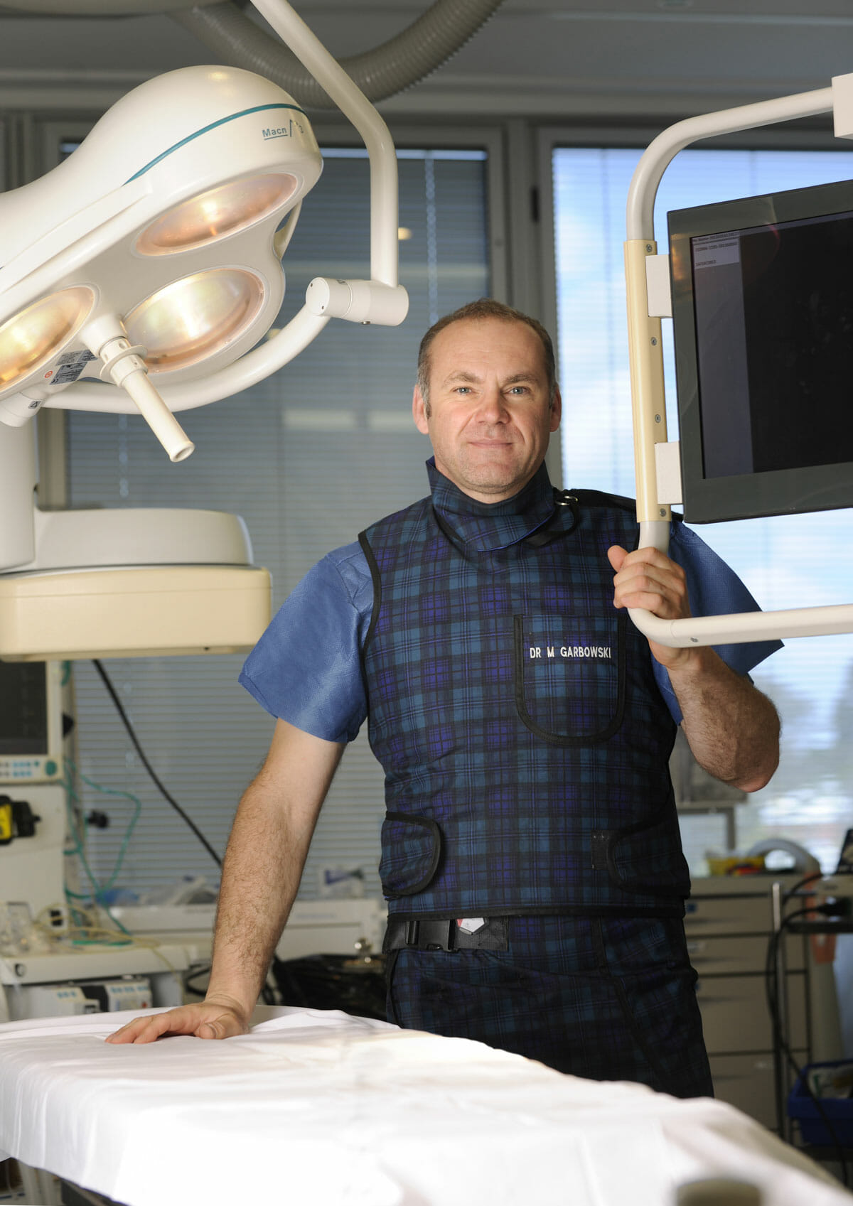
Dr Marek Garbowski has developed a strong clinical focus in the assessment and management of pelvic venous disorders, helping many women regain control of their lives after years of unexplained symptoms.
State-of-the-Art Assessment
Using the latest diagnostic imaging and vascular evaluation techniques, we ensure a thorough and accurate diagnosis. We work closely with gynaecologists and radiologists to deliver a multidisciplinary approach tailored to each patient’s needs.
Evidence-Based Treatment Options
Our clinic offers a full range of minimally invasive, image-guided treatment options — including embolisation of varicose pelvic veins, designed to relieve symptoms with minimal downtime.
Care for Public and Private Patients
Dr Garbowski provides consultations and procedures through both private practice and public hospital pathways, ensuring access to quality care for all patients.