
In brief
Spider veins (telangiectasias) and their slightly larger cousins, reticular veins, are among the most common vascular findings in adults. They are usually benign, but they can be cosmetically distressing, sometimes cause aching or burning, and occasionally signal underlying venous reflux that needs to be found and treated first. Good outcomes depend on assessing the whole venous system before treating the visible veins.
What they are
- Telangiectasias (spider veins): intradermal venules under 1 mm, red, blue or purple, in linear, arborising or “starburst” patterns. This is CEAP class C1.
- Reticular veins: blue-green subdermal veins of 1–3 mm. These often feed the telangiectasias above them.
How common, and why
Population studies such as the Edinburgh Vein Study found telangiectasias and reticular veins in a large majority of adults, and they are more frequent in women. Contributing factors include:
- Family history (strong genetic component)
- Female sex hormones, including pregnancy, the oral contraceptive pill and HRT
- Age, and prolonged standing occupations
- Sun-damaged skin and rosacea (facial telangiectasias)
- Previous trauma, surgery or sclerotherapy (matting)
- Underlying venous reflux, in a minority of patients
Presentation and examination
Most patients present with appearance concerns. Some report aching, heaviness, burning or itch, though the link between spider veins and symptoms is inconsistent, so look for another cause of the symptoms.
Examine the patient standing, with good lighting (a transilluminator or vein-light helps). Check for:
- Reticular feeder veins
- Varicose veins
- Corona phlebectatica at the ankle
- Oedema, pigmentation and lipodermatosclerosis
- Distribution: telangiectasias over the lateral thigh, buttock, vulva or perineum raise the possibility of a pelvic venous origin
Investigations
Ask for a venous duplex ultrasound in these cases:
- Symptoms out of proportion to the veins
- Visible varicose veins
- Signs of chronic venous disease
- A family history of significant venous disease
- Recurrence after treatment
- A distribution suggesting pelvic or perineal origin
A purely cosmetic C1 presentation without symptoms or signs does not always need imaging. A low threshold for duplex is reasonable in a specialist setting.
Management
Conservative. Graduated compression may relieve symptoms, but it does not remove spider veins. Sun protection, weight management and exercise help general venous health but will not clear existing veins.
Treat underlying reflux first. If duplex shows great or small saphenous or perforator reflux, treating the source (endovenous ablation, foam sclerotherapy or ambulatory phlebectomy as appropriate) reduces recurrence and improves the cosmetic result.
Definitive treatment of the visible veins
| Option | Best for | Notes |
|---|---|---|
| Liquid sclerotherapy (polidocanol or sodium tetradecyl sulfate) | Telangiectasias, small reticular veins | First-line for most patients. Very low concentrations are used for the finest veins. |
| Foam sclerotherapy | Reticular veins, larger feeders | Stronger effect, so use a small volume and take more care with visual and neurological symptoms. |
| Transcutaneous laser or IPL (Nd:YAG 1064 nm, pulsed dye, KTP) | Very fine vessels, needle-averse patients, matting, facial and ankle veins | Higher rates of pigmentary change and burns in darker skin types. Less effective for larger vessels. |
| Ambulatory phlebectomy | Larger reticular veins | Small incisions or punctures under local anaesthetic. |
| Combined (sclerotherapy plus laser) | Resistant or extensive disease | Treat feeders first, then the fine surface network. |
Practical points
- Expect 1-2 sessions, spaced 4–6 weeks apart depending upon number of veins
- Compression stockings after treatment, typically two weeks (daily application).
- Walking is encouraged; avoid intense exercise, heat and sun exposure for a period of 2 weeks after treatment.
- Complete clearance is not guaranteed. Most patients achieve substantial improvement, but new veins can appear over time and maintenance treatment is common.
- Treatment is usually cosmetic and generally not Medicare-rebatable unless there is symptomatic or significant venous disease requiring intervention.
When to wait: defer treatment in pregnancy (usually until after delivery and breastfeeding), acute DVT, active skin infection, known sclerosant allergy and severe immobility. Take extra care in patients with migraine with aura or a known right-to-left shunt.
Outcomes and natural history if untreated
Untreated spider veins are not dangerous. They tend to persist or slowly increase in number, especially with hormonal change and age. Bleeding is rare. The main clinical importance is as a marker of underlying reflux in the minority of patients who have it.
Complications and side effects
| Treatment | Common | Uncommon | Rare |
|---|---|---|---|
| Sclerotherapy | Stinging, cramping, bruising, urticarial wheals, temporary swelling | Hyperpigmentation 1-3% (usually fades over months, occasionally persists), telangiectatic matting, superficial thrombophlebitis or trapped blood | Skin ulceration or necrosis (extravasation, intra-arterial injection), visual disturbance or migraine, anaphylaxis, DVT, TIA or stroke (mainly foam) |
| Laser/IPL | Redness, swelling, purpura | Blistering, crusting, hyper- or hypopigmentation | Scarring, burns |
| Phlebectomy | Bruising, tenderness | Haematoma, hyperpigmentation | Nerve injury, infection |
Key points
- Most spider veins are benign and cosmetic; a focused venous history and examination will identify those that are not.
- Refer for duplex and specialist assessment if there are symptoms, varicose veins, skin changes, recurrence, or a pelvic or perineal distribution.
- Treating source reflux first improves outcomes.
- Counsel patients that treatment improves but does not “cure” the tendency to form new veins.
- Sclerotherapy is safe in experienced hands, but pigmentation and matting are the complications patients most often complain about, so warn them before they consent.
This page is general information and does not replace individual medical advice.

Listen to the Latest Podcast
Listen to the latest podcast featuring Dr Marek Garbowski and Medical Forum, WA’s independent monthly publication for health professionals.
Get in Touch
Treatments Available for Aesthetic Vascular Surgery – Spider Veins
Below are some of the treatments we have available for Aesthetic Vascular Surgery – Spider Veins. Click on the below to learn more or get in touch with us to request a consultation.
Ambulatory Phlebectomy
Ambulatory phlebectomy removes bulging varicose veins through very small punctures in the skin under local anaesthetic. It is a day procedure that leaves minimal scarring and allows a rapid return to normal activities.
Endovenous Laser Ablation (EVLT)
Endovenous laser ablation (EVLT) uses laser energy to close diseased veins from within, eliminating the underlying cause of varicose veins. It is a highly effective day procedure with a fast recovery and excellent long-term results.
Sclerotherapy
Sclerotherapy involves injecting a solution directly into varicose or spider veins to irritate and close the vessel wall. It is a quick, well-established outpatient treatment suitable for a range of vein sizes, from fine thread veins to medium-sized varicosities.
Why Choose Perth Vascular Clinic?
Expertise You Can Trust
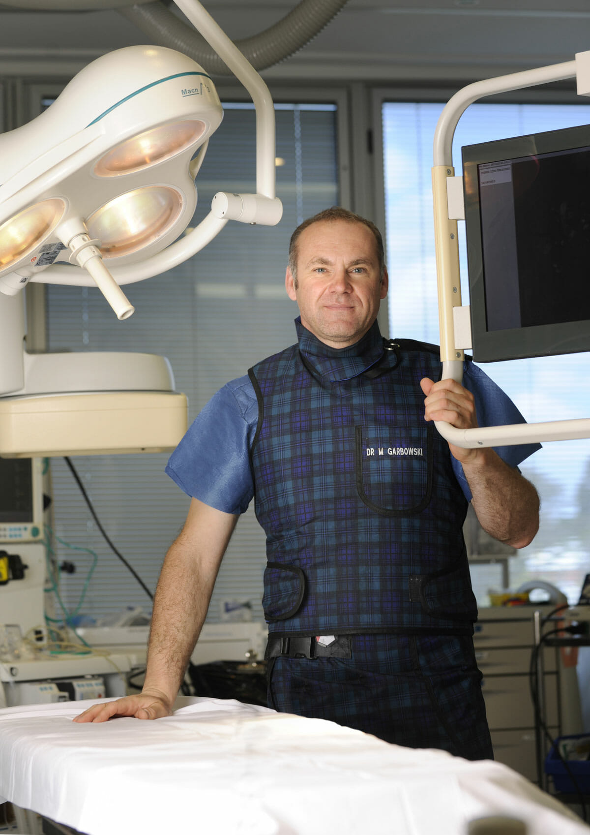
Dr Marek Garbowski has developed a strong clinical focus in the assessment and management of pelvic venous disorders, helping many women regain control of their lives after years of unexplained symptoms.
State-of-the-Art Assessment
Using the latest diagnostic imaging and vascular evaluation techniques, we ensure a thorough and accurate diagnosis. We work closely with gynaecologists and radiologists to deliver a multidisciplinary approach tailored to each patient’s needs.
Evidence-Based Treatment Options
Our clinic offers a full range of minimally invasive, image-guided treatment options — including embolisation of varicose pelvic veins, designed to relieve symptoms with minimal downtime.
Care for Public and Private Patients
Dr Garbowski provides consultations and procedures through both private practice and public hospital pathways, ensuring access to quality care for all patients.