
Overview
Thoracic outlet syndrome (TOS) describes a group of conditions caused by compression of the neurovascular structures (the brachial plexus, subclavian artery and subclavian vein) as they pass through the narrow corridor between the neck and the axilla known as the thoracic outlet. Depending on which structure is predominantly compressed, TOS is classified into three types: neurogenic (nTOS), venous (vTOS), and arterial (aTOS). Because symptoms can be vague and overlap with other cervical and shoulder conditions, TOS is frequently under-recognised and diagnosis is often delayed. Management ranges from targeted physiotherapy through to surgical decompression, and outcomes are generally favourable when the correct subtype is identified and treated appropriately.
Anatomy and Classification
The thoracic outlet is bounded by three potential sites of compression: the interscalene triangle (between the anterior and middle scalene muscles, with the first rib as its floor), the costoclavicular space (between the clavicle and first rib), and the retropectoralis minor space (beneath the pectoralis minor tendon). Compression at any of these levels can affect the brachial plexus, subclavian artery, or subclavian vein individually or in combination.
Thoracic outlet syndrome is divided into three clinical categories:
- Neurogenic TOS (nTOS): compression of the brachial plexus. This is by far the most common form, accounting for the large majority of cases.
- Venous TOS (vTOS): compression or thrombosis of the subclavian vein, classically presenting as effort-induced thrombosis (Paget-Schroetter syndrome).
- Arterial TOS (aTOS): compression of the subclavian artery, often associated with a cervical rib, which can lead to post stenotic dilatation, aneurysm formation, or distal embolisation.
A further descriptive category, “disputed” neurogenic TOS, is sometimes used for patients with clinical features suggestive of nerve compression but without objective electrodiagnostic confirmation.
Aetiology and Risk Factors
TOS results from a reduction in the available space within the thoracic outlet, or from repetitive strain across it. Common contributing factors include:
- Anatomical variants: a cervical rib, an elongated C7 transverse process, or an anomalous fibrous band, which narrow the interscalene triangle.
- Postural and musculoskeletal factors: scapular dyskinesia, rounded-shoulder posture, and pectoralis minor shortening, particularly from repetitive overhead activity.
- Trauma: whiplash injury, clavicle or first rib fracture, and repetitive occupational or sporting strain (common in swimmers, throwing athletes, weightlifters, and manual workers).
- Hypertrophy of the scalene or pectoralis minor muscles, seen in some overhead athletes and weightlifters.
- Congenital soft-tissue bands or a narrow costoclavicular space present from birth.
Neurogenic TOS is typically multifactorial, arising from a combination of anatomical predisposition and repetitive positional or postural strain on the brachial plexus. Venous TOS is usually related to a tight costoclavicular space combined with repetitive strenuous arm activity, while arterial TOS is most often driven by a bony abnormality such as a cervical rib.
Prevalence and Epidemiology
TOS is relatively uncommon, and reported incidence varies widely depending on diagnostic criteria used. Neurogenic TOS is the most prevalent subtype, accounting for most cases, while venous and arterial TOS are considerably rarer.
Estimates for neurogenic TOS suggest an incidence of roughly 2 to 3 cases per 100,000 people per year, with an overall prevalence of around 10 per 100,000. It typically affects adults in their 20s to 40s and is more common in women. Venous TOS tends to present in younger, otherwise healthy and often athletic individuals, reflecting its association with repetitive overhead or strenuous arm activity, while arterial TOS is rare and usually related to an underlying bony abnormality such as a cervical rib.
Clinical Presentation
Symptoms vary considerably depending on which structure is compressed, and often fluctuate with arm position or activity.
Neurogenic TOS presents with pain, paraesthesia, numbness, and weakness in the neck, shoulder, and arm, often radiating into the ulnar distribution of the hand. Symptoms are typically aggravated by overhead activity, carrying, or prolonged static postures, and may be associated with headache and a sense of arm heaviness.
Venous TOS presents with arm swelling, heaviness, cyanotic discolouration, and distended superficial collateral veins across the shoulder and chest wall, often appearing acutely after strenuous or repetitive arm use (Paget-Schroetter syndrome, or “effort thrombosis”).
Arterial TOS is the rarest and potentially most serious presentation, with symptoms of limb ischaemia: coldness, pallor, exercise-induced claudication of the arm, or, in more advanced cases, digital ischaemia or embolic phenomena from a post stenotic subclavian artery aneurysm.
Because symptoms can mimic cervical radiculopathy, peripheral nerve entrapment, and rotator cuff pathology, a careful history and examination, including provocative manoeuvres such as the Roos (elevated arm stress) test and Adson’s test, are essential to narrow the differential before proceeding to imaging.
Investigations
Diagnosis of TOS relies on a combination of clinical assessment and targeted imaging, guided by the suspected subtype.
- Plain radiography (cervicothoracic X-ray): first-line screening for a cervical rib, elongated C7 transverse process, or old clavicle/first rib fracture.
- Duplex ultrasound: dynamic scanning of the subclavian artery and vein with the arm in neutral and provocative (abducted/externally rotated) positions; particularly useful for venous TOS and for confirming positional compression.
- CT or MR angiography: defines bony and soft-tissue anatomy, vessel patency, aneurysmal change, and collateral formation; increasingly performed with the arm in both neutral and stress positions to demonstrate dynamic compression.
- Venography: remains the gold standard for confirming venous TOS and subclavian vein occlusion and is often performed as part of catheter-based treatment (thrombolysis or angioplasty).
- Nerve conduction studies and electromyography (EMG): used selectively in neurogenic TOS to exclude alternative or coexisting causes such as cervical radiculopathy or ulnar/median neuropathy; findings in nTOS are often normal or non-specific, so a normal study does not exclude the diagnosis.
- Diagnostic scalene block: an ultrasound-guided local anaesthetic (sometimes with botulinum toxin) injection into the anterior scalene muscle can support the diagnosis of neurogenic TOS when symptoms improve and may also guide treatment planning.
No single test is diagnostic for neurogenic TOS; the diagnosis remains primarily clinical, supported by imaging and, where relevant, a positive response to scalene block, with other causes of arm and neck symptoms carefully excluded.
Conservative Management
Conservative treatment is first line for the great majority of neurogenic TOS cases and forms the foundation of management even when surgery is ultimately required.
- Structured physiotherapy: the mainstay of treatment, focused on postural correction, scapular stabilisation, stretching of the scalene and pectoralis minor muscles, and nerve gliding exercises. A dedicated, TOS-specific physiotherapy program sustained over 6–12 weeks is generally required before benefit is assessed.
- Activity modification: avoiding or modifying provocative overhead or repetitive activities, particularly relevant for athletes and manual workers.
- Analgesia: simple analgesics and, where appropriate, neuropathic agents for nerve-related pain.
- Botulinum toxin injection: ultrasound-guided injection into the anterior scalene (and sometimes pectoralis minor) muscle can meaningfully reduce pain, typically for around two months or more, and is useful both as a therapeutic option and as a diagnostic aid in equivocal cases.
- Ergonomic and workplace modification: adjusting desk, driving, and sporting posture and technique to reduce mechanical strain on the thoracic outlet.
Most patients with neurogenic TOS improve with a sustained, well-directed conservative program, and surgery is generally reserved for those with persistent, functionally limiting symptoms despite an adequate trial of non-operative treatment, typically at least 6 months, unless red-flag features (progressive weakness, vascular compromise) mandate earlier surgical referral. Venous and arterial TOS, by contrast, more often require earlier intervention because of the risk of thrombosis, embolisation, or limb-threatening ischaemia.
Surgical Management
Surgery aims to decompress the thoracic outlet by removing or releasing the structures responsible for compression. The choice of approach depends on TOS subtype, anatomical findings, and surgeon experience.
Surgical approaches
- Transaxillary first rib resection: the traditional approach for neurogenic and venous TOS, performed through an incision in the axilla. It allows direct access to the first rib with resection of its middle portion, and is often combined with anterior and middle scalenectomy and excision of any fibrous bands or cervical rib.
- Supraclavicular approach: provides excellent exposure of the brachial plexus and subclavian artery, and is generally preferred when arterial reconstruction, cervical rib excision, or extensive neurolysis is required. First rib resection can be combined with this approach if needed.
- Rib-sparing scalenectomy: a newer, less invasive technique involving anterior and middle scalenectomy, brachial plexus neurolysis, and pectoralis minor release, without removing the first rib. Comparative data suggest similar 1-year functional and reintervention outcomes to first rib resection, with a lower complication profile, and it is increasingly favoured for neurogenic TOS in some centres.
- Robotic-assisted and minimally invasive first rib resection: emerging techniques allowing rib resection through smaller incisions with magnified visualisation; early single-centre series report outcomes comparable to open approaches, though longer-term and comparative data are still accumulating.
- Venous reconstruction: in venous TOS with chronic subclavian vein occlusion, first rib resection is often combined with venoplasty, and occasionally vein reconstruction or bypass, to restore patency after decompression.
- Arterial reconstruction: in arterial TOS with aneurysmal degeneration or thrombosis of the subclavian artery, rib and cervical rib resection is combined with arterial repair, patch angioplasty, or bypass grafting as required.
Perioperative pathway for venous TOS
Acute effort thrombosis of the subclavian vein is typically managed with catheter-directed thrombolysis or mechanical thrombectomy to restore vein patency, followed by surgical decompression (first rib resection with scalenectomy) once the acute thrombus has resolved, usually within a few weeks. Residual venous stenosis after decompression may require balloon venoplasty, sometimes performed as a staged procedure.
Postoperative course
Most patients undergo first rib resection as an inpatient procedure with a hospital stay of one to a few days. A structured post-operative physiotherapy program, generally beginning within the first one to two weeks, is important for restoring range of motion and preventing scar-related re-tethering of the brachial plexus. Return to full activity, including overhead and strenuous work, is typically staged over 6–12 weeks.
Complications and Side Effects
As with any surgery near major neurovascular structures, decompression for TOS carries specific risks that should be discussed with patients preoperatively.
- Pneumothorax: the pleural apex lies close to the operative field; this is one of the more common complications and is usually managed with a small chest drain if it occurs.
- Brachial plexus injury or neuropraxia: transient nerve irritation (paraesthesia, weakness) is relatively common in the early postoperative period and usually resolves over weeks to months; permanent nerve injury is uncommon but recognised.
- Injury to the long thoracic nerve or phrenic nerve: can cause scapular winging or hemidiaphragm elevation respectively; these are uncommon but recognised risks, particularly with the supraclavicular approach.
- Vascular injury: injury to the subclavian artery or vein during dissection is uncommon but can require intraoperative repair.
- Haematoma and wound infection: as with any surgical incision; haematoma in the axilla or supraclavicular region occasionally requires drainage.
- Chylothorax: rare, related to injury of the thoracic duct on the left side.
- Incomplete symptom relief or recurrence: particularly relevant to neurogenic TOS, where symptoms may persist or recur due to scar tissue formation (brachial plexus fibrosis), an incompletely resected first rib, or a missed contributing factor.
- Rib regrowth: rare, but can lead to recurrent compression if the rib is not adequately resected.
Across recent comparative series, complication rates for first rib resection and rib-sparing scalenectomy are broadly similar, in the order of 4–6%, with no significant difference in reintervention rates between the two techniques. Postoperative functional outcomes tend to be more favourable and more complete in venous and arterial TOS than in neurogenic TOS, likely because the compressive lesion can be more completely addressed in vascular TOS, whereas neurogenic symptoms depend on nerve recovery, which is inherently less predictable.
Long-Term Outcomes
Overall, surgical decompression for TOS is safe and effective for most appropriately selected patients, though the durability of benefit varies by subtype.
- Neurogenic TOS: reported success rates (good or excellent symptom relief) after first rib resection range widely across studies, generally in the order of 70–90% at intermediate follow-up, with somewhat lower rates of complete symptom resolution. Systematic review and meta-analysis data suggest transaxillary first rib excision, supraclavicular first rib excision with scalenectomy, and supraclavicular scalene release without rib resection produce broadly comparable success rates, though long-term functional outcomes remain less well characterised than short-term relief. A minority of patients experience symptom recurrence over subsequent years, often related to scar fibrosis or an incompletely addressed contributing factor.
- Venous TOS: outcomes after decompression combined with catheter-based treatment of the subclavian vein are generally excellent, with the majority of patients achieving durable symptom relief and vein patency, particularly when treated promptly after the acute thrombotic event. Outcomes are less favourable when there is chronic, long-standing venous occlusion prior to treatment.
- Arterial TOS: decompression with any necessary arterial reconstruction generally produces durable relief of ischaemic symptoms and removes the risk of further embolisation, reflecting the more complete correction of the underlying compressive lesion achievable in vascular TOS.
Across all subtypes, patient-reported quality of life and return to work or sport are strongly influenced by symptom duration prior to treatment, meaning earlier diagnosis and appropriately timed intervention tend to be associated with better long-term function. A structured, ongoing physiotherapy program following surgery further supports durable outcomes, particularly in neurogenic TOS.
Summary
Thoracic outlet syndrome is an uncommon but often under-recognised cause of arm, shoulder, and neck symptoms, arising from compression of the brachial plexus, subclavian artery, or subclavian vein within the thoracic outlet. Neurogenic TOS is the most common subtype and usually responds well to a structured, sustained physiotherapy program, with surgery reserved for those who do not improve. Venous and arterial TOS are less common but more often require earlier, combined medical, endovascular, and surgical treatment because of the risk of thrombosis, embolisation, or limb ischaemia.
Patients experiencing persistent arm, shoulder, or neck symptoms aggravated by overhead activity, or any swelling, discolouration, or coldness of the arm, should seek assessment. Early, accurate diagnosis, supported by targeted imaging and, where appropriate, specialist vascular surgical review, gives the best chance of a durable and complete recovery.
This information is provided for general educational purposes and does not replace individual clinical assessment. Please contact Perth Vascular Clinic to arrange a consultation to discuss your specific circumstances.

Listen to the Latest Podcast
Listen to the latest podcast featuring Dr Marek Garbowski and Medical Forum, WA’s independent monthly publication for health professionals.
Get in Touch
Treatments Available for Thoracic Outlet Syndromes
Below are some of the treatments we have available for Thoracic Outlet Syndromes. Click on the below to learn more or get in touch with us to request a consultation.
Why Choose Perth Vascular Clinic?
Expertise You Can Trust
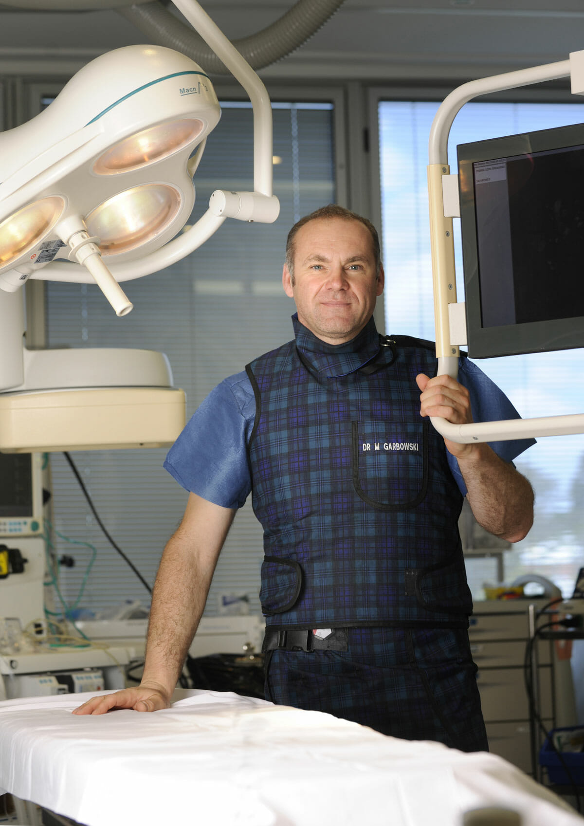
Dr Marek Garbowski has developed a strong clinical focus in the assessment and management of pelvic venous disorders, helping many women regain control of their lives after years of unexplained symptoms.
State-of-the-Art Assessment
Using the latest diagnostic imaging and vascular evaluation techniques, we ensure a thorough and accurate diagnosis. We work closely with gynaecologists and radiologists to deliver a multidisciplinary approach tailored to each patient’s needs.
Evidence-Based Treatment Options
Our clinic offers a full range of minimally invasive, image-guided treatment options — including embolisation of varicose pelvic veins, designed to relieve symptoms with minimal downtime.
Care for Public and Private Patients
Dr Garbowski provides consultations and procedures through both private practice and public hospital pathways, ensuring access to quality care for all patients.