
In brief
A deep vein thrombosis (DVT) is a blood clot in a deep vein, most often in the leg. Even with good treatment, many people are left with long-term leg problems. These can include swelling, heaviness, aching, skin changes and, in some cases, ulcers. This chronic condition is called post-thrombotic syndrome (PTS). It is not simply “a clot that never went away”. It is damage to the vein, and to the way blood flows through it, that persists after the clot has been treated. PTS is common, often under-recognised, and can substantially affect quality of life. Good options exist to reduce symptoms, protect the skin and, in selected patients, correct the underlying venous blockage.
What is chronic DVT and PTS?
After a DVT, the body breaks down the clot, but this is rarely complete. Two problems can persist:
- Obstruction: residual scar tissue or narrowing in the vein (most importantly in the iliac and femoral veins) blocks blood leaving the leg.
- Reflux: damage to the vein valves lets blood fall backwards down the leg.
Both cause ambulatory venous hypertension, where pressure in the leg veins stays high when standing or walking. This pressure overloads the small vessels and the calf muscle pump, and it drives the swelling, skin damage and ulceration of PTS. Chronic DVT describes the persistent post-thrombotic changes on imaging (residual thrombus, wall thickening, occlusion, collaterals). PTS is the clinical syndrome that results.
How common is it?
- About 20–50% of patients develop some degree of PTS after a symptomatic proximal DVT, despite adequate anticoagulation. Most cases declare themselves within the first two years.
- 5–10% develop severe PTS, and a smaller proportion develop venous ulcers.
- The risk is highest after iliofemoral DVT. In the control arm of the CaVenT trial, roughly 70% of patients treated with anticoagulation alone had PTS at 5 years.
- The impact on quality of life is comparable with other major chronic diseases.
Risk factors
- Proximal (femoral, iliofemoral) rather than calf DVT
- Ipsilateral recurrent DVT (roughly a six-fold increase in risk)
- Persistent leg symptoms one month after the DVT
- Obesity, older age, pre-existing varicose veins or chronic venous disease
- Sub-therapeutic anticoagulation in the early months, particularly with warfarin
- Residual thrombus or persistent iliac vein obstruction (including underlying May-Thurner anatomy)
Symptoms and signs
Symptoms: heaviness, aching, cramping, swelling, itching, pins and needles. These are typically worse at the end of the day and on prolonged standing or walking, and better with rest and elevation. Patients with iliofemoral obstruction may have venous claudication, a bursting pain in the thigh or calf on walking.
Signs:
- Pitting, then non-pitting, oedema
- Hyperpigmentation (haemosiderin staining), venous eczema, lipodermatosclerosis, atrophie blanche
- Secondary varicose veins and dilated collaterals over the groin, suprapubic area or abdominal wall
- Pain on calf compression
- Venous ulceration, typically over the medial malleolus
Diagnosis and severity grading
PTS is a clinical diagnosis, made no earlier than 3–6 months after the acute DVT, when the acute inflammatory symptoms have settled. The Villalta scale is the most widely used tool. It scores five symptoms and six signs from 0 (absent) to 3 (severe).
| Villalta score | Severity |
|---|---|
| 0–4 | No PTS |
| 5–9 | Mild PTS |
| 10–14 | Moderate PTS |
| ≥15, or venous ulcer | Severe PTS |
Other useful measures are the CEAP classification (2020 update), the Venous Clinical Severity Score (VCSS) and disease-specific quality-of-life tools such as VEINES-QOL.
Investigations
- Venous duplex ultrasound is the first-line test. It shows residual thrombus, wall thickening, loss of normal flow phasicity, collateral flow and valvular reflux (>1 second in the femoropopliteal segment, >0.5 second in the calf and profunda veins). It also excludes recurrent acute DVT.
- Ankle-brachial index should be measured before compression, to exclude significant arterial disease.
- CT or MR venography assesses the iliocaval segment when proximal obstruction is suspected.
- Venography with intravascular ultrasound (IVUS) is the reference standard for iliocaval obstruction and is more sensitive than venography alone. It is usually performed when intervention is being planned.
- Exclude other causes of leg swelling, including heart, kidney or liver disease, medications (for example calcium channel blockers), lymphoedema, lipoedema and arterial disease.
Prevention
- Effective anticoagulation for the acute DVT, with correct duration and adherence.
- Observational data and post hoc trial analyses suggest DOACs may be associated with a lower PTS risk than warfarin. This has not been confirmed in a dedicated randomised trial.
- Prompt mobilisation after DVT.
- Preventing recurrence, since recurrent ipsilateral DVT is the strongest modifiable risk factor.
- Compression stockings worn routinely after DVT do not prevent PTS (SOX trial) and are no longer recommended for that purpose. They remain useful for symptom relief.
- Early thrombus removal (catheter-directed thrombolysis or pharmacomechanical thrombectomy) may be considered in carefully selected patients with acute iliofemoral DVT, recent onset of symptoms, and low bleeding risk. In ATTRACT, it did not reduce overall PTS but reduced moderate-to-severe PTS, at the cost of more bleeding. CaVenT showed a reduction in PTS. Patient selection is critical.
Management of established PTS
Conservative measures (first line for all patients)
- Graduated compression stockings (typically 30–40 mmHg at the ankle) for symptom control, provided arterial supply is adequate. Evidence for benefit is modest but they remain a cornerstone of care. Intermittent pneumatic compression is an option for severe or refractory oedema.
- Exercise: structured walking and calf-strengthening exercises improve symptoms and calf pump function.
- Weight reduction, leg elevation and avoiding prolonged standing.
- Skin care: emollients for dry skin, topical corticosteroids for venous eczema, and early treatment of skin infection.
- Venoactive drugs (for example micronised purified flavonoid fraction) have modest evidence for symptom relief, and availability in Australia is variable. Diuretics are generally ineffective for venous oedema.
- Ulcer care: multilayer compression bandaging, wound care and treatment of the underlying venous hypertension.
Endovascular treatment
Venoplasty and stenting of iliofemoral or iliocaval obstruction is considered for patients with severe symptoms, or ulceration, despite optimal conservative therapy, when imaging or IVUS confirms significant outflow obstruction. Published series show meaningful improvement in pain and swelling and healing of a majority of ulcers. Patency is generally good in expert hands, although lower than for non-thrombotic stenoses. Adequate venous inflow (a patent femoral or profunda vein) improves stent patency. Post-procedure anticoagulation is standard and is often prolonged in post-thrombotic limbs.
Treatment of superficial venous reflux (endovenous ablation) can speed ulcer healing (EVRA trial). In PTS it should be done only after confirming that the superficial veins are not serving as essential collateral outflow for an obstructed deep system.
Open surgical treatment
Reserved for selected patients with severe disease when endovascular options are unsuitable or have failed:
- Femoro-femoral crossover bypass (Palma-Dale) for unilateral iliac vein occlusion
- Iliocaval or femoro-caval bypass, sometimes with a temporary arteriovenous fistula to improve flow
- Saphenopopliteal bypass (May-Husni) for femoral vein occlusion
- Common femoral vein endophlebectomy, usually as a hybrid with stenting
- Deep venous valve reconstruction (valvuloplasty, transposition, transplantation) for refractory deep reflux, which is technically demanding and performed in few centres
Outcomes and natural history without treatment
PTS is a chronic condition. Symptoms may stabilise or improve slowly with conservative care, but in a substantial minority they persist or progress. Untreated severe PTS can lead to recurrent, slow-healing venous ulcers, chronic skin changes, reduced mobility, cellulitis and lasting impairment of quality of life. Persistent iliofemoral obstruction and recurrent DVT are the main drivers of progression. Early recognition, consistent compression and exercise, and timely referral for assessment of venous outflow obstruction all improve the outlook.
Complications and side effects of treatment
| Treatment | Principal risks |
|---|---|
| Compression | Skin irritation, poor adherence, pressure injury if arterial disease is unrecognised |
| Catheter-directed thrombolysis or thrombectomy | Bleeding (including major bleeding), rarely intracranial haemorrhage, pulmonary embolism, contrast reaction, radiation exposure |
| Venoplasty and stenting | Stent thrombosis or restenosis, need for re-intervention, stent migration, groin haematoma, bleeding on anticoagulation |
| Open venous bypass | Graft occlusion, wound complications, infection, lymphatic leak, bleeding, and AVF-related swelling where a fistula is used |
| Valve reconstruction | Early failure or recurrent reflux, wound problems, DVT |
When to refer
- Persistent or worsening leg swelling and pain 3–6 months after DVT
- Venous ulceration, or skin changes progressing despite compression
- Venous claudication or prominent collateral veins
- Suspected recurrent DVT, or a suspected underlying iliac vein compression (May-Thurner) syndrome
- Moderate to severe PTS (Villalta ≥10) that is not controlled with conservative care
Key messages
- PTS affects up to half of patients after DVT and is more common and more severe after iliofemoral DVT.
- Prevention rests on effective anticoagulation and avoiding recurrence.
- Diagnose PTS clinically (Villalta), 3–6 months or more after the acute event.
- Compression, exercise and skin care are first line, but persistent severe symptoms justify assessment of iliofemoral outflow.
- Stenting can transform outcomes for suitable patients, and open surgery still has a role in complex disease.
Selected references
- Kahn SR, et al. Post-thrombotic syndrome: an AHA Scientific Statement. Circulation 2014;130:1636–61.
- Kahn SR, et al. Determinants and time course of the postthrombotic syndrome after acute deep venous thrombosis. Ann Intern Med 2008;149:698–707.
- Kahn SR, et al. Definition of post-thrombotic syndrome of the leg for use in clinical investigations: ISTH SSC. J Thromb Haemost 2009;7:879–83.
- Kahn SR, et al. Compression stockings to prevent post-thrombotic syndrome (SOX). Lancet 2014;383:880–8.
- Vedantham S, et al. Pharmacomechanical catheter-directed thrombolysis for DVT (ATTRACT). N Engl J Med 2017;377:2240–52.
- Enden T, et al. Catheter-directed thrombolysis in iliofemoral DVT (CaVenT). Lancet 2012;379:31–8. Long-term follow-up: Haig Y, et al. Lancet Haematol 2016;3:e64–71.
- Kakkos SK, et al. ESVS 2021 Clinical Practice Guidelines on the Management of Venous Thrombosis. Eur J Vasc Endovasc Surg 2021;61:9–82.
- Ortel TL, et al. ASH 2020 guidelines for management of VTE: treatment of DVT and PE. Blood Adv 2020;4:4693–738.
- Neglén P, et al. Stenting of the venous outflow in chronic venous disease. J Vasc Surg 2007;46:979–90.
- Gohel MS, et al. A randomized trial of early endovenous ablation in venous ulceration (EVRA). N Engl J Med 2018;378:2105–14.
- Lurie F, et al. The 2020 update of the CEAP classification system. J Vasc Surg Venous Lymphat Disord 2020;8:342–52.
- Kahn SR, Hirsch A, Shrier I. Effect of postthrombotic syndrome on health-related quality of life after DVT. Arch Intern Med 2002;162:1144–8.
This page is general information and does not replace individual medical advice.

Listen to the Latest Podcast
Listen to the latest podcast featuring Dr Marek Garbowski and Medical Forum, WA’s independent monthly publication for health professionals.
Get in Touch
Treatments Available for Chronic Deep Vein Thrombosis (Post Thrombotic Syndrome)
Below are some of the treatments we have available for Chronic Deep Vein Thrombosis (Post Thrombotic Syndrome). Click on the below to learn more or get in touch with us to request a consultation.
Angioplasty and Stenting
Angioplasty and stenting opens narrowed or blocked arteries by inflating a small balloon inside the vessel and, where needed, placing a mesh tube to hold it open. It is a minimally invasive alternative to open surgery for many arterial conditions.
Endovenous Laser Ablation (EVLT)
Endovenous laser ablation (EVLT) uses laser energy to close diseased veins from within, eliminating the underlying cause of varicose veins. It is a highly effective day procedure with a fast recovery and excellent long-term results.
Why Choose Perth Vascular Clinic?
Expertise You Can Trust
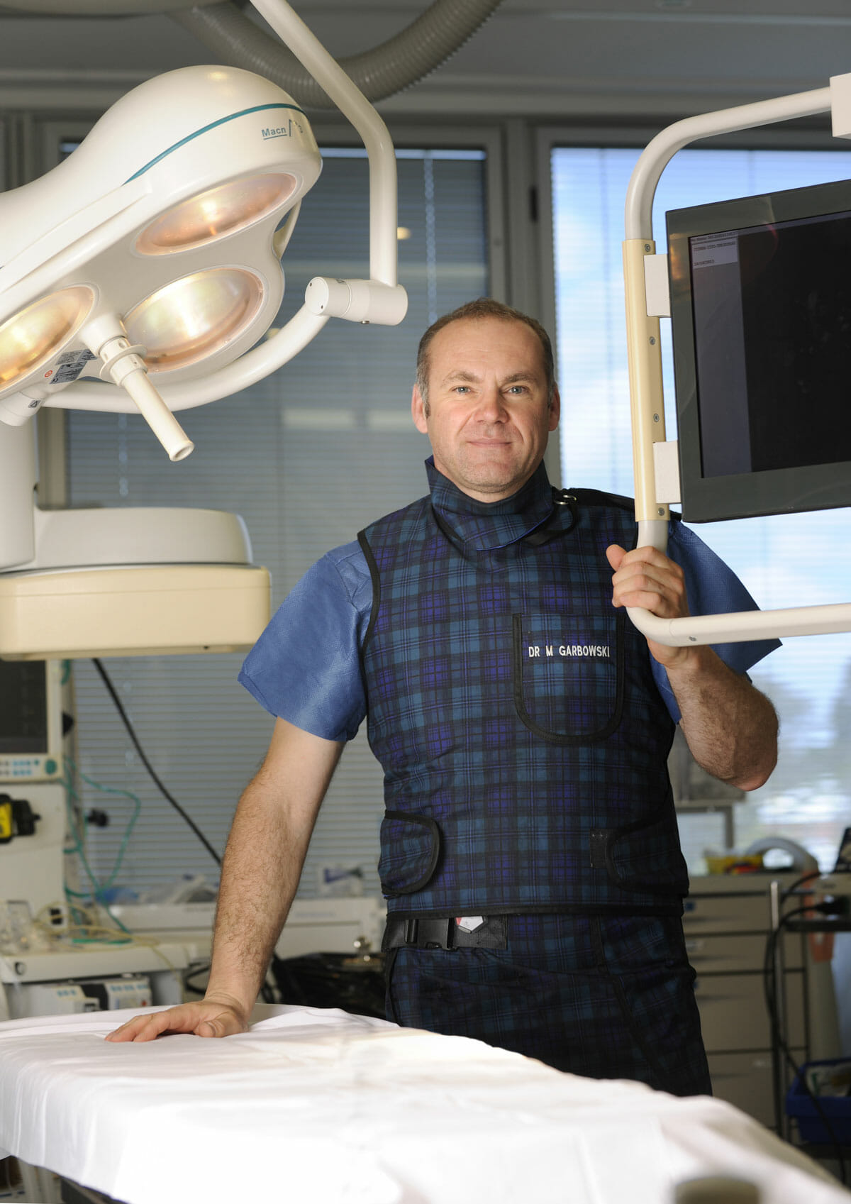
Dr Marek Garbowski has developed a strong clinical focus in the assessment and management of pelvic venous disorders, helping many women regain control of their lives after years of unexplained symptoms.
State-of-the-Art Assessment
Using the latest diagnostic imaging and vascular evaluation techniques, we ensure a thorough and accurate diagnosis. We work closely with gynaecologists and radiologists to deliver a multidisciplinary approach tailored to each patient’s needs.
Evidence-Based Treatment Options
Our clinic offers a full range of minimally invasive, image-guided treatment options — including embolisation of varicose pelvic veins, designed to relieve symptoms with minimal downtime.
Care for Public and Private Patients
Dr Garbowski provides consultations and procedures through both private practice and public hospital pathways, ensuring access to quality care for all patients.