
Overview
Acute superficial thrombophlebitis, also called superficial vein thrombosis (SVT), is a blood clot in a vein just beneath the skin, accompanied by inflammation of the vein wall. It is common, and it most often affects the legs. Although it was traditionally regarded as a minor, self-limiting problem, ultrasound studies have shown that SVT can extend into the deep venous system and is sometimes accompanied by deep vein thrombosis (DVT) or pulmonary embolism (PE). Careful assessment is therefore important.
Causes and risk factors
- Varicose veins, the most common setting, in which slow, disturbed flow favours clotting
- Previous SVT or venous thromboembolism (VTE)
- Pregnancy and the postpartum period, and oestrogen therapy (oral contraceptive pill, HRT)
- Obesity, older age, prolonged immobility, recent surgery or trauma
- Active cancer, or an inherited or acquired clotting tendency (thrombophilia)
- Intravenous cannulas, infusions or injections (the usual cause in the arm)
- Inflammatory conditions such as Behçet’s disease or Buerger’s disease (rare)
Clinical features
- A tender, warm, red, firm cord along the course of a superficial vein, often with surrounding skin redness
- Pain that is worse on standing or walking, with mild swelling around the vein
- Most often the great saphenous vein and its tributaries in the leg, less often the small saphenous vein
- Low-grade fever in some patients, though fever with spreading redness should raise concern about infection (suppurative thrombophlebitis, mainly cannula-related)
- Recurrent or migratory episodes in different veins, which may signal an underlying systemic cause such as malignancy or a thrombophilia
SVT is a clinical diagnosis but is easily confused with cellulitis, lymphangitis, erythema nodosum, panniculitis or DVT. Calf swelling or a swollen limb suggests a coexisting DVT.
Natural history
In most patients with a short segment of thrombosis, pain and tenderness settle over one to three weeks. The vein then becomes a firm, fibrous cord that may take months to soften, often leaving skin pigmentation. Recurrence is common when the underlying varicose veins are left untreated.
The main concern is clot propagation:
- Thrombus can extend along the vein towards the saphenofemoral junction (SFJ) in the groin or the saphenopopliteal junction (SPJ) behind the knee, and from there into the deep veins.
- In large registry data (the POST study), about one in four patients with lower limb SVT had a coexisting DVT at diagnosis, and around 1 in 20 had symptomatic PE.
- Of patients without DVT at presentation, a minority developed symptomatic VTE or clot extension in the following weeks when not anticoagulated.
The risk is greatest with longer segments of thrombus (5 cm or more), thrombus close to the SFJ or SPJ, involvement of the main saphenous trunks above the knee, and additional risk factors such as prior VTE, cancer or thrombophilia.
Investigations
Duplex ultrasound is the key investigation. It is recommended for lower limb SVT to:
- confirm the diagnosis and exclude cellulitis or other causes
- identify which vein is involved (great saphenous, small saphenous or a tributary) and measure the length of thrombus
- measure the distance of the thrombus from the SFJ or SPJ
- exclude concurrent DVT in both legs
- map the underlying venous reflux and varicose veins for later treatment
Blood tests are not routinely needed. D-dimer is unhelpful because it is elevated by the inflammation itself. Inflammatory markers and cultures are used if infection is suspected. Thrombophilia testing and cancer screening should be reserved for recurrent, migratory, unprovoked or unusual-site SVT, or for patients with a suggestive personal or family history. Cancer screening should follow age-appropriate guidelines, not extensive imaging.
Management
Treatment depends on the ultrasound findings and the patient’s risk profile.
| Ultrasound finding | Usual approach |
|---|---|
| Concurrent DVT | Treat as DVT with full therapeutic anticoagulation |
| Thrombus within 3 cm of SFJ/SPJ, or extending into the deep system | Therapeutic anticoagulation, generally for at least 3 months, with vascular specialist review |
| Thrombus 5 cm or longer, more than 3 cm from the junction | Prophylactic-dose anticoagulation for about 45 days (fondaparinux 2.5 mg daily, prophylactic LMWH, or rivaroxaban 10 mg daily) |
| Short segment (under 5 cm), below the knee, away from junctions | Symptomatic treatment; consider anticoagulation if risk factors are present; repeat ultrasound at about 7 to 10 days to detect extension |
The dose, agent and duration should be individualised, and some of these uses are off-label.
Symptomatic measures (all patients)
- Regular walking and avoiding prolonged immobility
- Graduated compression stockings or bandaging, as tolerated
- Warm compresses, and leg elevation at rest
- Topical NSAIDs, or a short course of oral NSAIDs in patients without contraindications, for pain and inflammation
Anticoagulation
The CALISTO trial showed that fondaparinux 2.5 mg daily for 45 days reduced symptomatic VTE, clot extension and recurrence by more than 80% compared with placebo. The SURPRISE trial found oral rivaroxaban 10 mg daily to be a comparable alternative. Guidelines (ESVS 2021, CHEST 2021) support these approaches. Pregnancy calls for LMWH, and DOACs are avoided.
Procedural options
- Office-based evacuation of clot from a thrombosed varicose tributary can give rapid pain relief in selected patients.
- Treating the underlying varicose disease, by endovenous ablation, foam sclerotherapy or phlebectomy, is usually planned once the acute inflammation has settled, to reduce the risk of recurrence.
- Open ligation of the SFJ is now rarely required and is reserved for selected cases.
Special situations
- Cannula-related SVT: remove the cannula; treat with topical therapy and elevation. Suppurative thrombophlebitis needs antibiotics and sometimes surgical excision.
- Recurrent or migratory SVT: investigate for an underlying systemic cause.
- Hormonal therapy: review the need for oestrogen-containing medication.
When to seek urgent medical attention
Seek prompt review if you develop:
- swelling of the whole leg, or calf pain and tenderness
- chest pain, breathlessness, or coughing up blood
- rapidly spreading redness, fever or pus
- a tender cord extending towards the groin or behind the knee
Key sources
- Kakkos SK et al. ESVS 2021 Clinical Practice Guidelines on the management of venous thrombosis. Eur J Vasc Endovasc Surg 2021
- Stevens SM et al. Antithrombotic therapy for VTE disease: second update of the CHEST guideline. Chest 2021
- Decousus H et al. Fondaparinux for the treatment of superficial-vein thrombosis in the legs (CALISTO). N Engl J Med 2010
- Decousus H et al. Superficial venous thrombosis and venous thromboembolism: a large, prospective epidemiologic study (POST). Ann Intern Med 2010
- Beyer-Westendorf J et al. Rivaroxaban versus fondaparinux for SVT (SURPRISE). Lancet Haematol 2017
This page is general information and does not replace individual medical advice.

Listen to the Latest Podcast
Listen to the latest podcast featuring Dr Marek Garbowski and Medical Forum, WA’s independent monthly publication for health professionals.
Get in Touch
Treatments Available for Acute Superficial Thrombophlebitis
Below are some of the treatments we have available for Acute Superficial Thrombophlebitis. Click on the below to learn more or get in touch with us to request a consultation.
Ambulatory Phlebectomy
Ambulatory phlebectomy removes bulging varicose veins through very small punctures in the skin under local anaesthetic. It is a day procedure that leaves minimal scarring and allows a rapid return to normal activities.
Endovenous Laser Ablation (EVLT)
Endovenous laser ablation (EVLT) uses laser energy to close diseased veins from within, eliminating the underlying cause of varicose veins. It is a highly effective day procedure with a fast recovery and excellent long-term results.
Radiofrequency Ablation (RFA)
Radiofrequency ablation uses precisely controlled heat to seal diseased veins from the inside, causing them to close and be gradually absorbed by the body. It is a highly effective, walk-in walk-out treatment for varicose veins with minimal discomfort and downtime.
Why Choose Perth Vascular Clinic?
Expertise You Can Trust
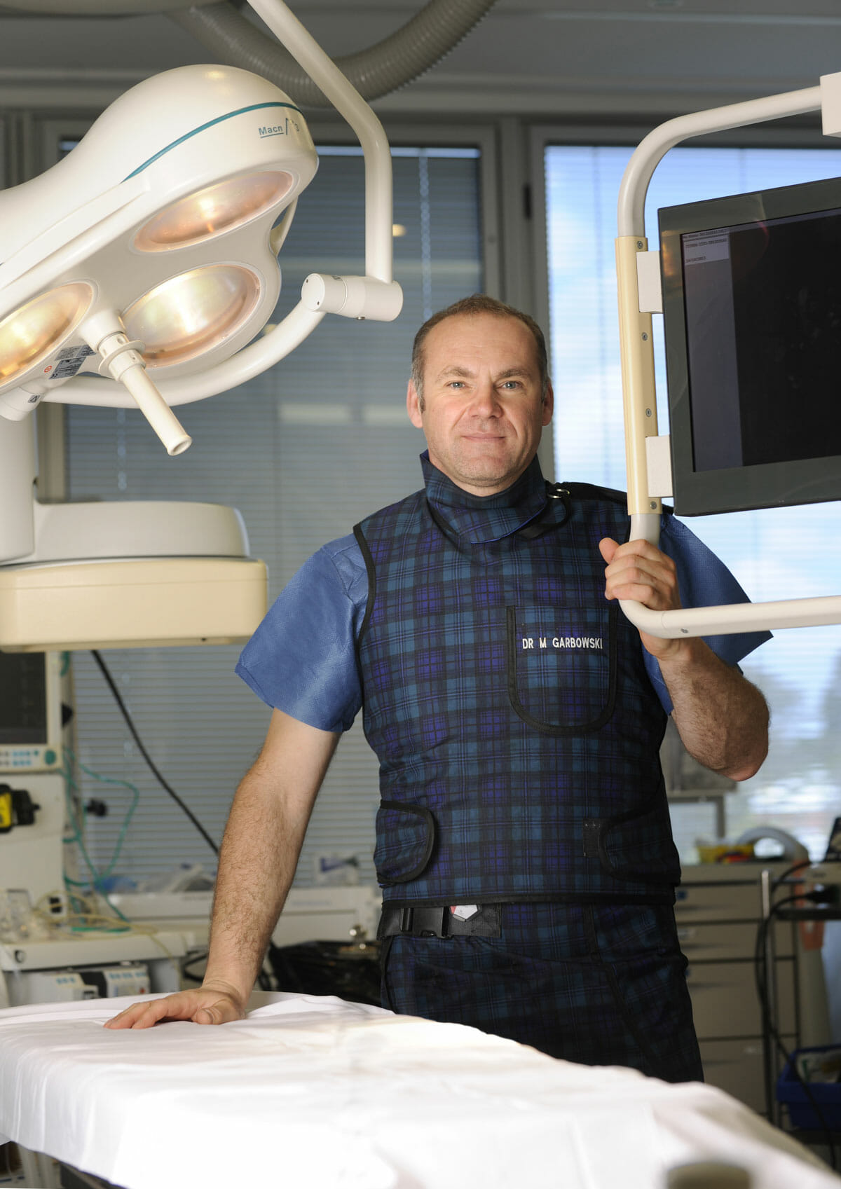
Dr Marek Garbowski has developed a strong clinical focus in the assessment and management of pelvic venous disorders, helping many women regain control of their lives after years of unexplained symptoms.
State-of-the-Art Assessment
Using the latest diagnostic imaging and vascular evaluation techniques, we ensure a thorough and accurate diagnosis. We work closely with gynaecologists and radiologists to deliver a multidisciplinary approach tailored to each patient’s needs.
Evidence-Based Treatment Options
Our clinic offers a full range of minimally invasive, image-guided treatment options — including embolisation of varicose pelvic veins, designed to relieve symptoms with minimal downtime.
Care for Public and Private Patients
Dr Garbowski provides consultations and procedures through both private practice and public hospital pathways, ensuring access to quality care for all patients.