
Overview
Arterial ulcers are wounds caused by poor blood supply to the leg or foot. An arterial (ischaemic) ulcer develops when a limb does not receive enough blood flow to keep its tissue alive. The usual cause is peripheral arterial disease (PAD), a narrowing or blockage of the arteries supplying the leg, almost always due to atherosclerosis. Once blood flow falls below the level the skin needs to survive minor everyday trauma, even a small knock, blister or tight shoe can break down into a wound that will not close on its own.
Unlike most leg ulcers, arterial ulcers will not heal with dressings and compression alone. The underlying blockage in the artery has to be addressed first. This is different from the far more common venous leg ulcer, which arises from valve failure and pooling of blood in the veins rather than a lack of arterial supply. Correctly telling the two apart matters clinically, because the standard treatment for venous ulcers, firm compression bandaging, can be harmful and even limb-threatening if applied to a leg with unrecognised arterial disease.
Prevalence
Peripheral arterial disease becomes increasingly common with age, affecting an estimated one in ten to fifteen people over 65, and a substantially higher proportion of those who smoke or have diabetes. Chronic leg ulcers of any cause affect roughly 1 to 2% of adults over 60.
Of all chronic leg ulcers, venous disease accounts for the majority (around 60 to 70%), with arterial disease responsible for a further 10 to 15% and mixed arterial/venous disease for much of the remainder. Arterial ulcers are therefore less common than venous ulcers overall, but disproportionately serious: they carry a far higher risk of progressing to gangrene and amputation if the underlying arterial blockage is not treated.
Causes
The dominant cause is atherosclerosis, the gradual build-up of plaque inside the artery wall, narrowing the vessels that supply the leg and foot. The main drivers of this process are well established and mostly modifiable:
- Smoking: the single strongest modifiable risk factor for peripheral arterial disease
- Diabetes mellitus: accelerates atherosclerosis and often affects smaller, more distal vessels
- Hypertension and dyslipidaemia (high blood pressure and abnormal cholesterol)
- Chronic kidney disease
- Advancing age and family history of vascular disease
The ulcer itself is usually triggered by a minor, often trivial, injury such as a knock, an ill-fitting shoe or a scratch, which in a normally perfused leg would heal without incident but in an ischaemic leg cannot. Pressure areas such as the toes, heel and lateral shin are particularly vulnerable.
Signs and Symptoms
Arterial ulcers have a fairly distinctive appearance and history, which helps distinguish them from venous ulcers at the bedside:
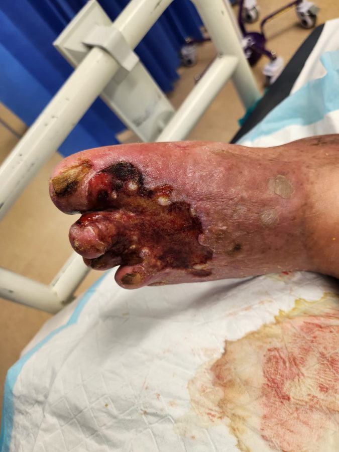
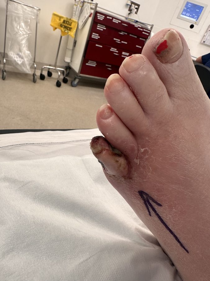
- Location: toes, heel, lateral malleolus (outer ankle), shin and other pressure points, rather than the inner gaiter area typical of venous ulcers
- Edge: well-defined and “punched out”
- Base: pale, grey or necrotic, and dry, with minimal fluid
- Pain: often severe, worse at night, and eased by hanging the leg down over the side of the bed
- Surrounding skin: shiny, hairless, cool and pale
- Pulses: reduced or absent at the ankle and foot
Other supporting signs of significant PAD include dependent rubor (redness when the leg hangs down) with pallor on elevation, delayed capillary refill, and a history of intermittent claudication (cramping pain on walking) or rest pain in the calf or foot.
Arterial ulcer or venous ulcer?
| Feature | Arterial ulcer | Venous ulcer |
|---|---|---|
| Location | Toes, heel, lateral malleolus, shin, pressure points | Medial gaiter area (above the inner ankle) |
| Edge | Well-defined, “punched out” | Irregular, sloping |
| Base | Pale, grey or necrotic; dry | Pink/red granulation; moist |
| Exudate | Minimal | Often moderate to heavy |
| Pain | Often severe, worse at night, eased by hanging the leg down | Usually mild, eased by elevation |
| Surrounding skin | Shiny, hairless, cool, pale | Pigmented, eczematous, warm |
| Pulses | Reduced or absent | Usually palpable |
What arterial ulcers look like
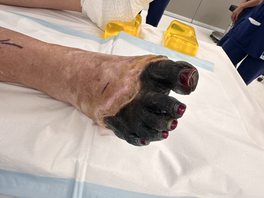
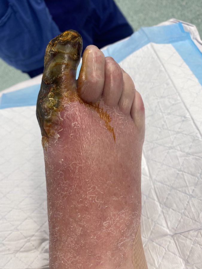
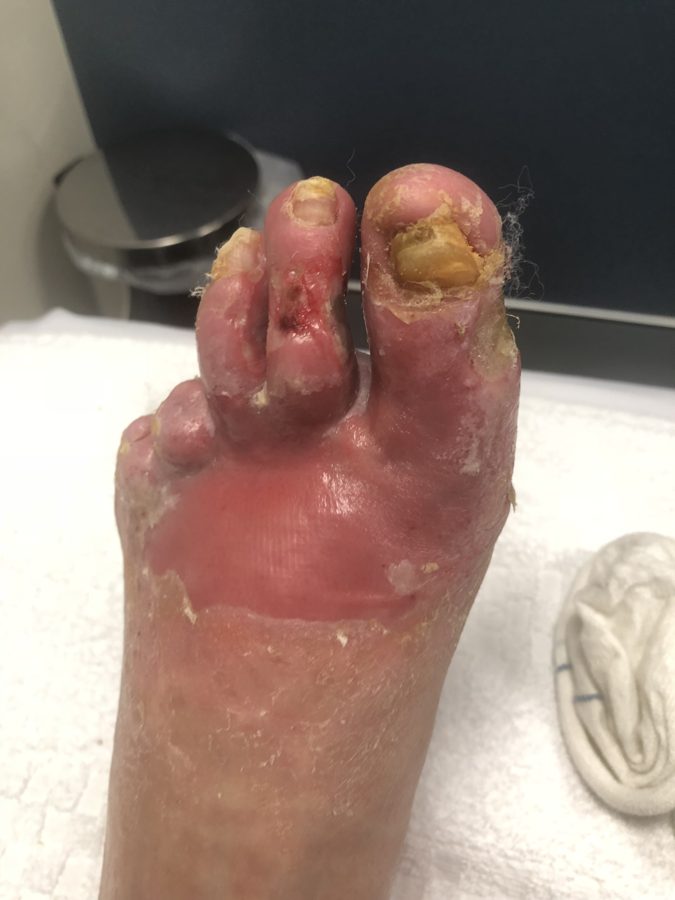
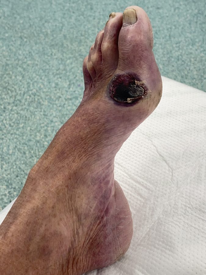
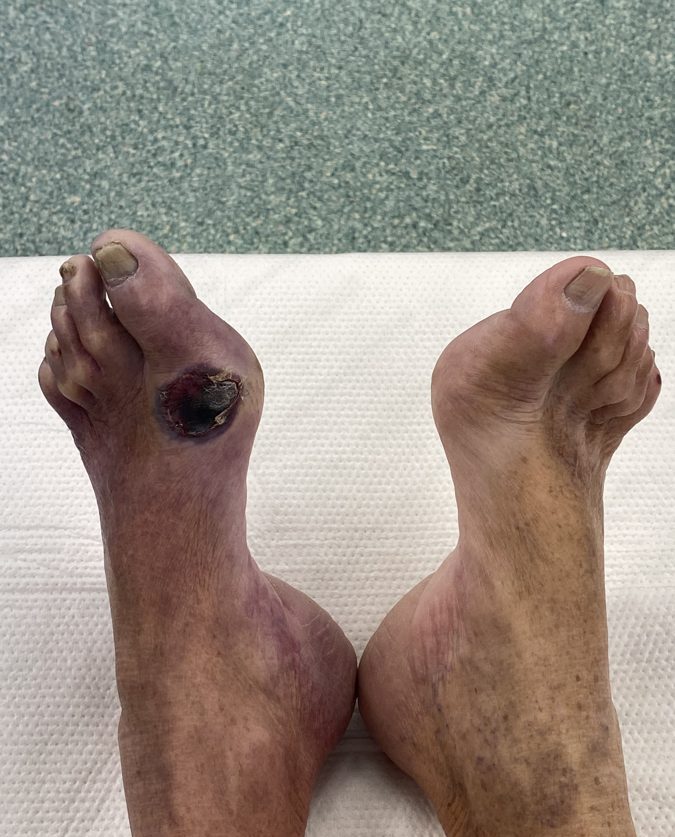
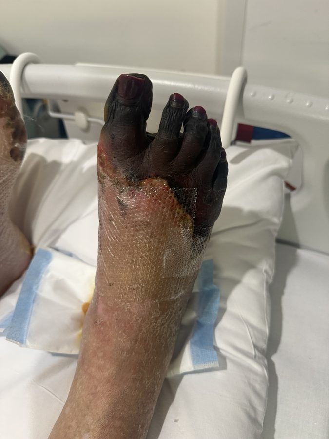
The images below show real examples of arterial ulceration and the tissue loss that follows when blood supply to the foot is critically reduced, from early ischaemic ulcers of the toes through to established gangrene. Click any image to view it at full size.
Complications of Untreated Arterial Ulcers
Arterial ulcers do not heal spontaneously while the underlying blood supply remains inadequate. Left untreated, they tend to enlarge, become infected and progress toward gangrene. This trajectory, known as chronic limb-threatening ischaemia, carries a substantial risk of major amputation within a year if flow is not restored. Patients with PAD severe enough to cause tissue loss also carry a markedly increased risk of heart attack and stroke, reflecting the systemic nature of atherosclerosis.
Early recognition and referral materially change this trajectory: the sooner blood flow is restored, the better the chance of healing and limb salvage.
Assessment and Management at Perth Vascular Clinic
Investigations
Assessment aims to confirm the diagnosis, quantify how severe the arterial insufficiency is, and map the disease for treatment planning.
At first assessment
Pulse examination: femoral, popliteal, posterior tibial and dorsalis pedis pulses.
Ankle-brachial pressure index (ABPI): the ratio of ankle to arm systolic pressure, performed in our ABI Clinic. Values below 0.9 suggest PAD, and below 0.5 suggest severe disease. ABPI can be falsely elevated in diabetes or renal disease due to arterial calcification, in which case a toe-brachial index is more reliable.
Imaging
Arterial duplex ultrasound: non-invasive first-line mapping of narrowings and blockages.
CT angiography: a detailed anatomical roadmap from the aorta to the foot, widely used for planning.
MR angiography: an alternative where CT contrast is contraindicated.
Digital subtraction angiography: invasive catheter-based imaging, usually reserved for the time of intervention.
Supporting work-up
- HbA1c, fasting lipids and renal function.
- Wound swab and inflammatory markers if infection is suspected.
- Cardiovascular risk assessment, as PAD and coronary or cerebrovascular disease frequently coexist.
Treatment options
Successful management rests on three pillars: modifying the underlying risk factors, restoring arterial blood flow, and caring for the wound itself. Wound care and compression are supportive at best and cannot substitute for revascularisation. Compression, in particular, should not be applied until adequate arterial supply has been confirmed.
Medical and supportive management
Smoking cessation: the single most impactful modifiable step.
Optimised glycaemic control in diabetes.
Statin and antiplatelet therapy for cardiovascular risk reduction.
Blood pressure control.
Structured wound care, debridement, infection control and offloading of pressure areas.
Supervised exercise therapy where claudication coexists and revascularisation is not immediately required.
Endovascular treatment
Catheter-based techniques, including balloon angioplasty, stenting, drug-coated balloons and atherectomy for heavily calcified disease, are used to open narrowed or blocked segments from within the vessel. They are performed through a small puncture, usually under local anaesthetic, with a shorter recovery than open surgery. Endovascular treatment suits focal or moderate-length disease and higher-risk patients well, though long occlusions treated this way can be less durable and may need repeat intervention over time.
Open surgical treatment
Bypass grafting (using the patient’s own vein or a prosthetic conduit) reroutes blood flow around a blocked segment, while endarterectomy surgically removes plaque from a focally diseased artery, most often at the groin. Hybrid procedures combine both approaches in a single setting. Open surgery is generally favoured for longer-segment occlusive disease, after failed endovascular treatment, or where the anatomy and the patient’s fitness favour a durable surgical repair.
The choice between endovascular and open repair, or a staged combination of both, depends on the pattern and length of disease, the quality of the vessels available as a target, and the patient’s overall fitness for surgery. This decision is individualised and made in conjunction with the vascular surgical team.
Multidisciplinary wound care
Once flow is restored, healing is supported by a team that typically includes the vascular surgeon, a wound care nurse, podiatry (particularly for diabetic patients) and, for larger defects, plastic surgery input for skin grafting or flap reconstruction.
For general practitioners: when to refer
Refer promptly to a vascular surgeon for any leg or foot ulcer where arterial disease is suspected, including:
- Absent or markedly reduced pedal pulses
- Rest pain, particularly nocturnal pain relieved by hanging the leg over the bed
- An ulcer that has not progressed toward healing within two weeks of appropriate wound care
- Any suggestion of gangrene, spreading infection, or a diabetic foot ulcer
- An ABPI below 0.9, or an unreliable or falsely elevated ABPI in a diabetic or dialysis patient
Hold off on compression bandaging until arterial sufficiency has been confirmed. Inappropriate compression in an ischaemic limb can precipitate rapid deterioration.
For patients: key points
- A leg wound that is painful, slow to heal, or associated with cold or pale skin should be checked promptly rather than managed with dressings alone.
- Arterial ulcers heal once blood flow is restored. The wound itself is a symptom of the underlying blockage, not a separate problem.
- Stopping smoking and controlling blood sugar, blood pressure and cholesterol all improve both healing and long-term outcomes.
- Treatment is tailored to the individual. Many patients are suitable for minimally invasive endovascular treatment, while others benefit more from open surgical bypass.
Patients can be referred to Perth Vascular Clinic for assessment of any leg or foot ulcer where arterial disease is suspected, or for any concerning leg wound where the underlying cause is uncertain.
This information is provided for general educational purposes and does not replace individual medical assessment. Please consult your GP or a vascular specialist for advice specific to your circumstances.

Listen to the Latest Podcast
Listen to the latest podcast featuring Dr Marek Garbowski and Medical Forum, WA’s independent monthly publication for health professionals.
Get in Touch
Treatments Available for Arterial Ulcers
Below are some of the treatments we have available for Arterial Ulcers. Click on the below to learn more or get in touch with us to request a consultation.
Angioplasty and Stenting
Angioplasty and stenting opens narrowed or blocked arteries by inflating a small balloon inside the vessel and, where needed, placing a mesh tube to hold it open. It is a minimally invasive alternative to open surgery for many arterial conditions.
Bypass Surgery
Bypass surgery creates a new route for blood to travel around a blocked or severely narrowed artery. It is used to restore circulation to the legs and feet when less invasive treatments are not suitable or have not been effective.
Why Choose Perth Vascular Clinic?
Expertise You Can Trust
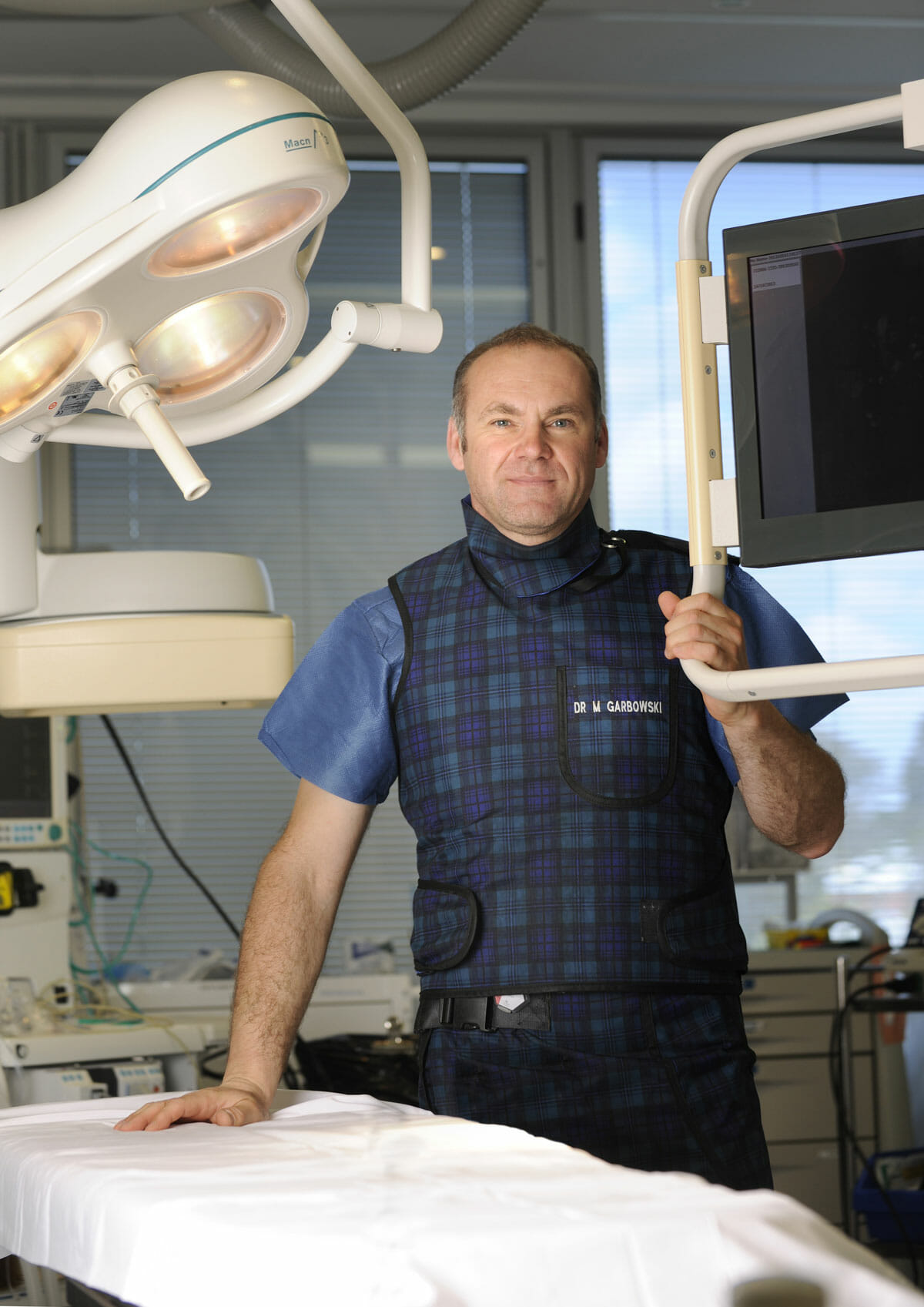
Dr Marek Garbowski has developed a strong clinical focus in the assessment and management of pelvic venous disorders, helping many women regain control of their lives after years of unexplained symptoms.
State-of-the-Art Assessment
Using the latest diagnostic imaging and vascular evaluation techniques, we ensure a thorough and accurate diagnosis. We work closely with gynaecologists and radiologists to deliver a multidisciplinary approach tailored to each patient’s needs.
Evidence-Based Treatment Options
Our clinic offers a full range of minimally invasive, image-guided treatment options — including embolisation of varicose pelvic veins, designed to relieve symptoms with minimal downtime.
Care for Public and Private Patients
Dr Garbowski provides consultations and procedures through both private practice and public hospital pathways, ensuring access to quality care for all patients.
Frequently Asked Questions
An arterial (ischaemic) ulcer is a wound on the leg or foot that develops because the limb is not receiving enough blood flow to keep its tissue alive. The usual cause is peripheral arterial disease, a narrowing or blockage of the leg arteries due to atherosclerosis. Even a minor knock or a tight shoe can break down into a wound that will not close on its own.