
Overview
Blood vessels usually travel through the body in protected channels. In a few places, however, an artery, vein or hollow organ must pass through a narrow gap between fixed structures such as bone, ligament, muscle or another vessel. If that gap is too tight, or the tissues around it are abnormal, the vessel can be squeezed, causing a vascular compression syndrome.
Veins have thin walls and low internal pressure, so they flatten easily, and compression leads to congestion, swelling, pain and clotting. Arteries are more resistant, but repeated compression can injure the wall. This can lead to narrowing, blockage, aneurysm formation or clots that travel downstream. In one condition, superior mesenteric artery syndrome, it is the arteries that compress a segment of bowel. Compression syndromes are often overlooked because symptoms can be vague and imaging is usually normal at rest.
Venous compression syndromes
Nutcracker syndrome (left renal vein compression)
The left renal vein travels from the left kidney across the midline to reach the inferior vena cava. On its way it passes through a narrow space between two arteries: the superior mesenteric artery (SMA) in front and the abdominal aorta behind. The SMA leaves the aorta at an angle, and the fat pad and lymphatic tissue that fill this space normally hold the SMA away from the aorta. The angle is typically 38° to 65°.
When that angle narrows (commonly below about 25° to 30°), the vein is squeezed like a nut in a nutcracker. This is more likely in people with low body weight, rapid weight loss, or an exaggerated lumbar curve. Less commonly, a retroaortic left renal vein is compressed between the aorta and the vertebral body (posterior nutcracker).
Pressure builds in the kidney side of the vein and is relieved through alternative routes. These include the left gonadal (ovarian or testicular) vein, pelvic veins and ureteric veins. The consequences are:
- flank or pelvic pain
- blood in the urine
- protein in the urine when upright
- a left varicocele in men
- pelvic venous congestion in women
May-Thurner syndrome (iliac vein compression)
The two common iliac veins join at the level of the fifth lumbar vertebra to form the inferior vena cava. The right common iliac artery crosses in front of the left common iliac vein here, pressing it against the lumbar spine behind. Over many years the constant pulsation can also cause scar-like webs or spurs to form inside the vein.
Because of this anatomy, the left leg is affected far more often than the right. Presentations include:
- persistent left leg swelling
- heaviness and aching
- varicose veins
- venous ulceration in advanced disease
- deep vein thrombosis (DVT), sometimes extensive and without an obvious trigger
Compression of the iliac veins can also contribute to pelvic venous disorders.
Venous thoracic outlet syndrome (Paget-Schroetter syndrome)
The thoracic outlet is the passage between the neck and the arm, and it has three compartments where compression can occur.
- Interscalene triangle: the gap between the anterior and middle scalene muscles and the first rib. The subclavian artery and brachial plexus pass through it.
- Costoclavicular space: the gap between the collarbone, the first rib and the subclavius muscle. The subclavian vein runs here, in front of the anterior scalene muscle and separate from the artery and nerves, so it is the most confined.
- Retropectoral space: the space beneath the pectoralis minor tendon.
In venous TOS the subclavian vein is repeatedly compressed in the costoclavicular space, particularly during overhead arm activity. This can cause sudden clotting (effort thrombosis) in young, active people. It presents with swelling, blue-purple discolouration and a heavy, aching arm.
Arterial compression syndromes
Median arcuate ligament syndrome (celiac artery compression)
The median arcuate ligament is a fibrous band that joins the two sides of the diaphragm as it wraps around the aorta, at about the level of the twelfth thoracic vertebra. In most people it lies above the origin of the celiac artery, the first major branch supplying the stomach, liver and spleen. In some people it sits lower and presses on the artery, and the celiac nerve tissue nearby can be involved as well.
Compression increases when breathing out, giving a characteristic hooked appearance of the artery on side-on imaging. Symptoms include:
- upper abdominal pain, often after eating
- nausea
- weight loss
- fear of eating
The condition is a diagnosis of exclusion, because other causes of these symptoms are far more common.
Popliteal artery entrapment syndrome
Normally the popliteal artery passes behind the knee, medial to and beneath the medial head of the gastrocnemius (calf) muscle. In popliteal entrapment, the artery follows an unusual course or the muscle has an abnormal attachment, so the artery is squeezed when the calf contracts. Fibrous bands or the popliteus muscle can be responsible. In some people there is no anatomical abnormality and the muscle is simply enlarged (functional entrapment).
It typically affects young, athletic people, causing calf cramping on exertion. Repeated compression can lead to arterial narrowing, a post-stenotic aneurysm, blockage, or small clots that travel to the foot.
Arterial thoracic outlet syndrome
This is the least common form of thoracic outlet syndrome, but potentially the most serious. It is usually associated with a cervical rib or an abnormal first rib pressing on the subclavian artery in the interscalene triangle. The artery can become narrowed, then dilated (aneurysmal), and can throw clots into the arm and hand. This can cause coldness, pain, colour change or, in severe cases, tissue loss in the fingers.
Compression of the bowel by arteries
Superior mesenteric artery (SMA) syndrome (duodenal compression)
The same narrow gap between the SMA and the aorta that holds the left renal vein also holds the third (horizontal) part of the duodenum. The duodenum crosses the midline at about the level of the third lumbar vertebra, below the left renal vein. It passes in front of the aorta and inferior vena cava and behind the SMA, and it is held in this position by the ligament of Treitz at its upper end. The mesenteric fat pad that surrounds the SMA keeps the artery away from the aorta and gives the duodenum room to pass.
When this space narrows, the duodenum is pinned between the two vessels and food cannot pass through. The aortomesenteric angle falls below about 22° to 25° (normally 38° to 65°), and the distance between the artery and the aorta falls below about 8 mm (normally 10 to 28 mm). The usual cause is loss of the protective fat pad, for example from:
- significant or rapid weight loss
- severe burns or trauma
- prolonged bed rest
- malabsorption
- restrictive eating disorders
- bariatric surgery
- surgical correction of scoliosis, or a body cast that stretches the spine
Anatomical variants such as an abnormally low origin of the SMA or a short ligament of Treitz make it more likely.
Symptoms are those of a partial high bowel obstruction:
- upper abdominal pain and fullness after eating
- early satiety and nausea
- vomiting, which may contain bile
- weight loss
Symptoms often ease when lying prone, on the left side, or in the knee-chest position, because these positions widen the aortomesenteric angle. Because weight loss both causes and results from the condition, it can become a self-perpetuating cycle. Complications include dehydration, electrolyte disturbance, malnutrition, and enlargement of the stomach and proximal duodenum.
Diagnosis rests on CT or MR angiography, which can measure the angle and distance directly and show a dilated duodenum ending abruptly at the point of compression. A barium or contrast study shows the same obstruction functionally. Other causes of duodenal obstruction must be excluded, and SMA syndrome can occur together with nutcracker syndrome.
Treatment is usually conservative first, and it succeeds in most people. It focuses on restoring nutrition and body weight, using small frequent meals, positioning strategies, and where necessary nasojejunal or parenteral feeding. If this fails, or in severe or long-standing cases, surgical options include duodenojejunostomy (bypassing the compressed segment, often laparoscopically), gastrojejunostomy, or mobilisation of the duodenum by dividing the ligament of Treitz.
Summary of anatomy
| Syndrome | Structure compressed | Structures compressing it | Typical features |
|---|---|---|---|
| Nutcracker | Left renal vein | SMA and aorta | Haematuria, flank/pelvic pain |
| May-Thurner | Left common iliac vein | Right common iliac artery against L5 | Left leg swelling, DVT |
| Venous TOS | Subclavian vein | First rib, clavicle, subclavius | Arm swelling, effort thrombosis |
| MALS | Celiac artery | Median arcuate ligament | Post-meal pain, weight loss |
| Popliteal entrapment | Popliteal artery | Gastrocnemius or fibrous bands | Calf claudication in young adults |
| Arterial TOS | Subclavian artery | Cervical or first rib | Cold, painful hand; emboli |
| SMA syndrome | Third part of duodenum | SMA and aorta | Post-meal pain, vomiting, weight loss |
Diagnosis
The starting point is a detailed history and examination, including provocative positions where relevant (arm elevation, ankle flexion, respiration). Duplex ultrasound is the first-line test for vascular syndromes and can assess flow dynamically. CT or MR angiography and venography can show the anatomy in detail. Catheter-based venography with intravascular ultrasound (IVUS) and pressure measurements can confirm whether a compression is haemodynamically significant.
Treatment
Treatment depends on the syndrome, the severity of symptoms and any complications. Options include:
- Observation and conservative management, including compression therapy, anticoagulation when there is thrombosis, weight or activity modification, and nutritional restoration in SMA syndrome
- Endovascular treatment, such as thrombolysis, angioplasty and stenting, most established for iliac vein compression
- Surgical decompression, such as first rib resection for TOS, release of the median arcuate ligament, or popliteal release with arterial reconstruction when the artery is damaged
- Bowel bypass (duodenojejunostomy) for SMA syndrome that does not respond to conservative measures
When to seek specialist advice
Consider a vascular assessment if you have unexplained one-sided leg or arm swelling, recurrent or unprovoked clots, exertional limb pain in a young and otherwise healthy person, persistent pelvic or flank pain with blood in the urine, or post-meal abdominal pain with weight loss, especially with vomiting. Early diagnosis can prevent complications.
This article is general information and is not a substitute for individual medical advice.

Listen to the Latest Podcast
Listen to the latest podcast featuring Dr Marek Garbowski and Medical Forum, WA’s independent monthly publication for health professionals.
Get in Touch
Treatments Available for Arterial and Venous Compression Syndromes
Below are some of the treatments we have available for Arterial and Venous Compression Syndromes. Click on the below to learn more or get in touch with us to request a consultation.
Why Choose Perth Vascular Clinic?
Expertise You Can Trust
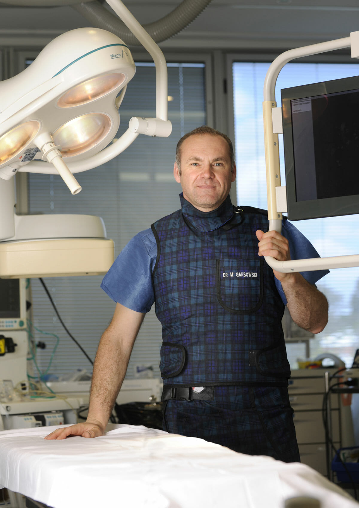
Dr Marek Garbowski has developed a strong clinical focus in the assessment and management of pelvic venous disorders, helping many women regain control of their lives after years of unexplained symptoms.
State-of-the-Art Assessment
Using the latest diagnostic imaging and vascular evaluation techniques, we ensure a thorough and accurate diagnosis. We work closely with gynaecologists and radiologists to deliver a multidisciplinary approach tailored to each patient’s needs.
Evidence-Based Treatment Options
Our clinic offers a full range of minimally invasive, image-guided treatment options — including embolisation of varicose pelvic veins, designed to relieve symptoms with minimal downtime.
Care for Public and Private Patients
Dr Garbowski provides consultations and procedures through both private practice and public hospital pathways, ensuring access to quality care for all patients.