
Overview
Venous leg ulcers are open wounds on the lower leg that arise from chronically elevated pressure in the veins of the leg (venous hypertension), most commonly due to incompetent (leaking) valves in the superficial or deep venous system. They are the most common type of leg ulcer, accounting for the large majority of chronic lower limb ulceration seen in general practice, wound clinics and vascular surgery. Unlike simple wounds, venous ulcers are a manifestation of underlying chronic venous disease and rarely heal permanently unless the underlying venous abnormality is identified and treated.
Prevalence
Chronic venous disease is extremely common, affecting a substantial proportion of the adult population, with visible varicose veins present in around a fifth to a third of adults and more advanced skin changes in a smaller subset. Venous leg ulcers specifically affect roughly 1% of adults at some point in their lives, with prevalence rising sharply with age. Active ulceration is seen in approximately 1 to 3% of people over 65. Venous disease is responsible for 60 to 80% of all chronic leg ulcers, making it far more common than arterial, diabetic or mixed-aetiology ulcers.
Risk factors include increasing age, female sex, obesity, prolonged standing occupations, previous deep vein thrombosis (DVT), family history of varicose veins, multiple pregnancies, and previous leg trauma or fracture. Venous ulcers have a significant tendency to recur. Up to 70% recur within five years without adequate treatment of the underlying venous incompetence, making correct diagnosis and definitive management essential rather than optimistic wound dressing alone.
Causes
Venous ulceration develops through a common final pathway of venous hypertension: sustained elevation of pressure within the venous system of the leg, particularly during standing and walking when pressure should normally fall due to the calf muscle pump. The mechanisms behind this include:
- Superficial venous reflux: incompetence of the great or small saphenous veins, or their major tributaries, allowing blood to reflux downward instead of being returned to the heart. This is the single most common and most treatable cause.
- Deep venous reflux or obstruction: valve failure in the femoral or popliteal veins, or obstruction from previous DVT (post-thrombotic syndrome), which is harder to correct but still amenable to some interventions.
- Perforator vein incompetence: failed one-way valves in the small veins connecting superficial and deep systems, allowing high pressure to be transmitted directly to the skin and subcutaneous tissue.
- Calf muscle pump failure: reduced ankle mobility, immobility or neuromuscular disease reducing the effectiveness of the calf as a venous pump.
- Pelvic venous disease: reflux from incompetent ovarian or internal iliac veins, or iliac vein compression (May-Thurner syndrome), can contribute to lower limb venous hypertension, particularly in women, and is often under-recognised as a driver of atypical or recurrent venous ulceration.
Sustained venous hypertension leads to capillary damage, leakage of fibrinogen and red cell breakdown products into the tissue, chronic inflammation, and progressive skin and subcutaneous fibrosis (lipodermatosclerosis). This fragile, poorly nourished skin eventually breaks down, often after minor trauma, to form an ulcer that fails to heal because the driving venous hypertension persists.
Signs and Symptoms
Venous ulcers typically occur over the medial gaiter area of the lower leg (the region above the inner ankle bone), though lateral and less commonly circumferential ulcers occur. Typical features include:
- Irregular but shallow ulcer edges, with a moist, granulating or fibrinous base
- Surrounding haemosiderin staining (brownish skin discolouration from chronic red cell breakdown)
- Lipodermatosclerosis: firm, woody induration and narrowing of the lower calf (an “inverted champagne bottle” leg shape)
- Atrophie blanche: pale, scarred, star-shaped areas of skin
- Venous eczema (varicose eczema) with itching, scaling and weeping skin
- Visible varicose veins or telangiectasia
- Ankle and lower leg swelling, worse at the end of the day and improved with elevation
- Aching, heaviness or cramping in the leg, typically relieved by elevation and worsened by prolonged standing
- Pulses are usually palpable and normal (an important distinguishing feature from arterial ulcers) unless there is coexisting peripheral arterial disease
Pain is often present but is typically less severe than in arterial ulcers, and characteristically improves with leg elevation, the opposite pattern to arterial disease, where elevation worsens pain.
What venous ulcers look like
The images below show real examples of venous ulcers and the skin changes that accompany them, from active ulceration through to healed scarring. Click any image to view it at full size.


Complications of Untreated Venous Ulcers
Leaving venous ulcers untreated, or managing them with wound dressings alone without addressing the underlying venous hypertension, carries significant risks:
- Chronic non-healing and recurrence: ulcers may persist for months to years, or heal and recur repeatedly, without correction of venous reflux.
- Infection: cellulitis, spreading soft tissue infection and, in severe cases, osteomyelitis of underlying bone.
- Increasing wound size and depth, occasionally exposing tendon or bone.
- Malignant transformation (Marjolin’s ulcer): squamous cell carcinoma arising within a chronic, long-standing ulcer, an uncommon but serious complication requiring biopsy of any atypical or non-healing ulcer edge.
- Progressive lipodermatosclerosis and limb fibrosis, causing permanent leg shape change and reduced mobility.
- Significant impact on quality of life: chronic pain, exudate, odour, impaired mobility, sleep disturbance, social isolation and depression are well documented in patients with long-standing venous ulcers.
- Substantial healthcare and economic burden: chronic wound care, dressings and nursing visits over years, with associated costs to the patient and health system.
- Sepsis, in severe or neglected infected ulcers, occasionally requiring hospitalisation.
Early, correct diagnosis and treatment of the underlying venous abnormality, rather than ulcer dressings in isolation, is central to preventing these outcomes.
Assessment and Management at Perth Vascular Clinic
Perth Vascular Clinic offers a comprehensive, structured pathway for the assessment and treatment of venous leg ulcers, combining specialist vascular surgical expertise with in-house diagnostic imaging and the full range of modern conservative, endovenous and surgical treatment options.
Comprehensive assessment
- Specialist vascular consultation with a fellowship-trained vascular and endovascular surgeon, including a detailed history and examination of the ulcer, leg and vascular system.
- In-house diagnostic vascular ultrasound, including duplex mapping of the superficial, deep and perforator venous systems to precisely identify the source and extent of venous reflux or obstruction.
- Ankle-brachial pressure index (ABPI) and arterial assessment in our ABI Clinic, to safely exclude or identify coexisting arterial disease before compression therapy is commenced.
- CEAP staging of chronic venous disease to guide and document treatment planning.
- Assessment for pelvic venous contributors to lower limb venous disease in appropriate patients, including referral for pelvic venous imaging where indicated.
- Wound assessment, including biopsy referral where an atypical or non-healing ulcer raises concern for an alternative diagnosis.
Management options
Perth Vascular Clinic provides the full spectrum of modern venous ulcer management under one roof, tailored to each patient’s individual venous anatomy and general health:
- Conservative therapy: supervised compression bandaging and stockings through our Compression Stockings Clinic, wound care advice, and coordination with wound care nursing and community services.
- Endovenous ablation: radiofrequency ablation (RFA), endovenous laser ablation and other minimally invasive techniques to close incompetent superficial veins under local anaesthetic, as an office-based procedure.
- Ultrasound-guided foam sclerotherapy for residual varicosities and perforator incompetence.
- Open venous surgery, including saphenous vein ligation and stripping, for selected patients where this remains the most appropriate option.
- Iliac vein stenting for patients with significant iliac venous outflow obstruction contributing to non-healing or recurrent ulceration.
- Ovarian and internal iliac vein embolisation for patients in whom pelvic venous reflux is contributing to lower limb venous disease.
- Coordinated multidisciplinary care, liaising with GPs, wound care nurses and other specialists to optimise healing and prevent recurrence.
- Structured follow-up, including surveillance duplex ultrasound, to confirm treatment success and monitor for recurrence.
Patients can be referred to Perth Vascular Clinic for assessment of a non-healing, recurrent or newly diagnosed venous leg ulcer, or for any concerning leg wound where the underlying cause is uncertain.
This information is provided for general educational purposes and does not replace individual medical assessment. Please consult your GP or a vascular specialist for advice specific to your circumstances.

Listen to the Latest Podcast
Listen to the latest podcast featuring Dr Marek Garbowski and Medical Forum, WA’s independent monthly publication for health professionals.
Get in Touch
Treatments Available for Venous Leg Ulcers
Below are some of the treatments we have available for Venous Leg Ulcers. Click on the below to learn more or get in touch with us to request a consultation.
Ambulatory Phlebectomy
Ambulatory phlebectomy removes bulging varicose veins through very small punctures in the skin under local anaesthetic. It is a day procedure that leaves minimal scarring and allows a rapid return to normal activities.
Endovenous Laser Ablation (EVLT)
Endovenous laser ablation (EVLT) uses laser energy to close diseased veins from within, eliminating the underlying cause of varicose veins. It is a highly effective day procedure with a fast recovery and excellent long-term results.
Non-invasive Assessment of PVD – ABI Clinic
Our ABI Clinic uses a simple, painless ankle-brachial pressure test to measure blood flow in your legs and screen for peripheral arterial disease. Results guide your treatment plan with no invasive procedures required.
Pelvic Vein Embolisation
Pelvic vein embolisation is a minimally invasive procedure that blocks the enlarged, poorly functioning veins causing pelvic congestion syndrome. It is performed under image guidance with no general anaesthetic required and provides lasting relief from chronic pelvic pain.
Radiofrequency Ablation (RFA)
Radiofrequency ablation uses precisely controlled heat to seal diseased veins from the inside, causing them to close and be gradually absorbed by the body. It is a highly effective, walk-in walk-out treatment for varicose veins with minimal discomfort and downtime.
Sclerotherapy
Sclerotherapy involves injecting a solution directly into varicose or spider veins to irritate and close the vessel wall. It is a quick, well-established outpatient treatment suitable for a range of vein sizes, from fine thread veins to medium-sized varicosities.
Stockings Clinic
Our Stockings Clinic provides professional measurement and fitting of graduated compression stockings to help manage varicose veins, chronic leg swelling, and venous ulcers. Correctly fitted stockings make a significant difference to comfort and outcomes.
VARIXIO Foam Sclerotherapy
VARIXIO foam sclerotherapy is a clinically proven, minimally invasive treatment for varicose veins using licensed polidocanol injectable foam to close affected veins and restore healthy leg circulation.
Why Choose Perth Vascular Clinic?
Expertise You Can Trust
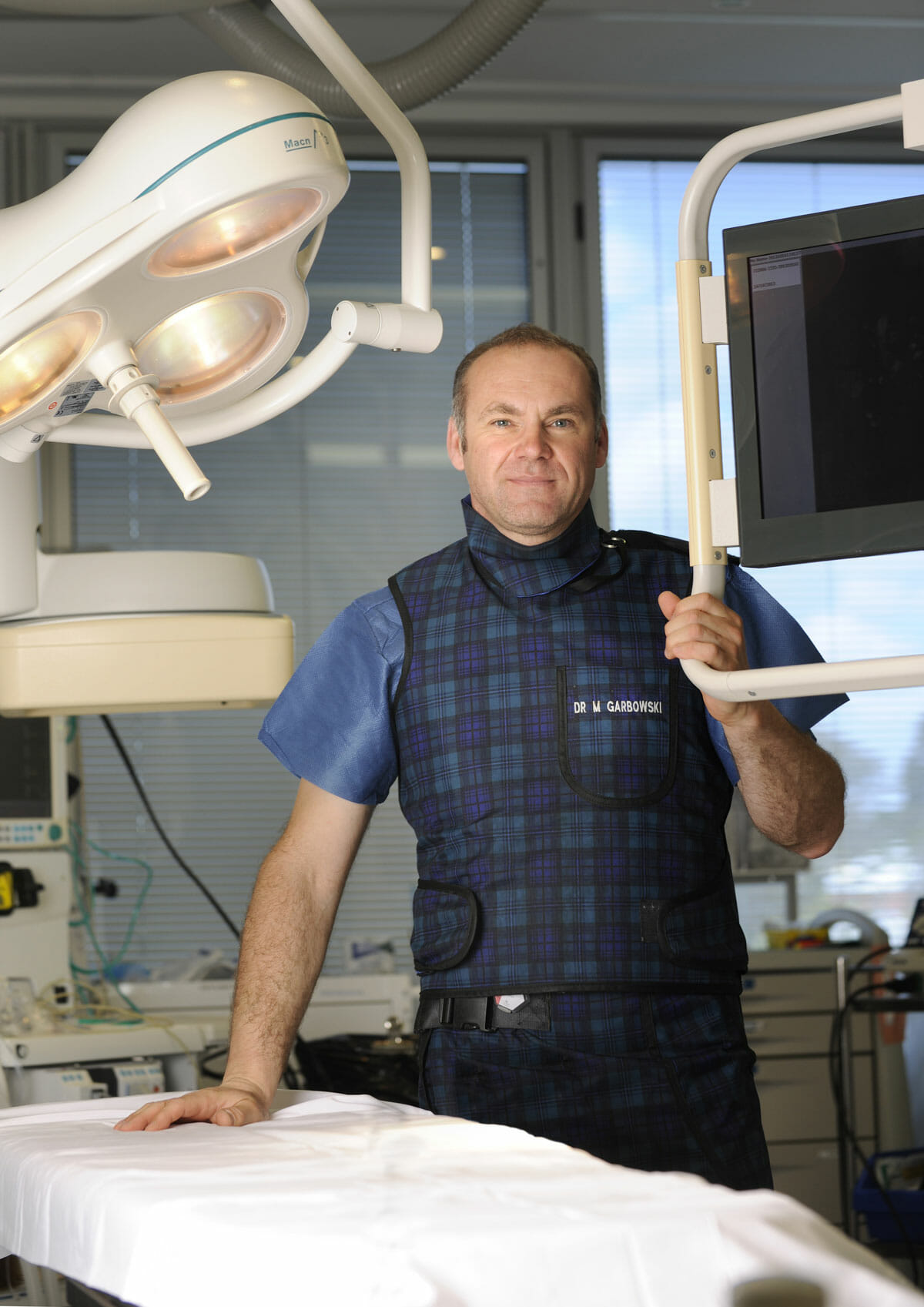
Dr Marek Garbowski has developed a strong clinical focus in the assessment and management of pelvic venous disorders, helping many women regain control of their lives after years of unexplained symptoms.
State-of-the-Art Assessment
Using the latest diagnostic imaging and vascular evaluation techniques, we ensure a thorough and accurate diagnosis. We work closely with gynaecologists and radiologists to deliver a multidisciplinary approach tailored to each patient’s needs.
Evidence-Based Treatment Options
Our clinic offers a full range of minimally invasive, image-guided treatment options — including embolisation of varicose pelvic veins, designed to relieve symptoms with minimal downtime.
Care for Public and Private Patients
Dr Garbowski provides consultations and procedures through both private practice and public hospital pathways, ensuring access to quality care for all patients.
Frequently Asked Questions
A venous leg ulcer is an open wound on the lower leg, usually near the inner ankle, caused by long-standing high pressure in the leg veins (venous hypertension). It is the most common type of leg ulcer and is a sign of underlying chronic venous disease rather than a simple skin injury.