
Why narrowing of the neck arteries matters for stroke, how it is assessed, and how it is treated: medical therapy, carotid endarterectomy, carotid stenting and TCAR. Written for patients and referring doctors.
In this article
In brief
- Carotid artery disease is narrowing of the arteries that carry blood to the brain, usually from atherosclerosis. It matters because of stroke: fragments of plaque or clot can break off and lodge in the brain or the eye.
- The most important question is whether the narrowing has caused symptoms in the last six months. After a recent TIA, transient loss of vision in one eye or minor stroke, a 70–99% stenosis carries a high early risk of stroke, and surgery works best when it is done within two weeks.
- For people without symptoms, best medical therapy is the foundation of care. The CREST-2 trials (2025) found that adding stenting to intensive medical therapy reduced stroke, while adding endarterectomy did not do so significantly, so procedures are now offered selectively.
- There are three ways to open a narrowed carotid artery: endarterectomy (CEA), transfemoral stenting (CAS) and transcarotid artery revascularisation (TCAR). CEA remains the reference standard. The best choice depends on symptoms, timing, anatomy, age and other health conditions.
- A suspected TIA or stroke is an emergency. It should be assessed the same day, not through a routine outpatient queue.
Call 000 straight away for sudden weakness or numbness of the face, arm or leg, trouble speaking or understanding, or loss of vision in one eye, even if it passes within minutes. A short episode is a warning, and the risk of a stroke is highest in the first days.
What is carotid artery disease?
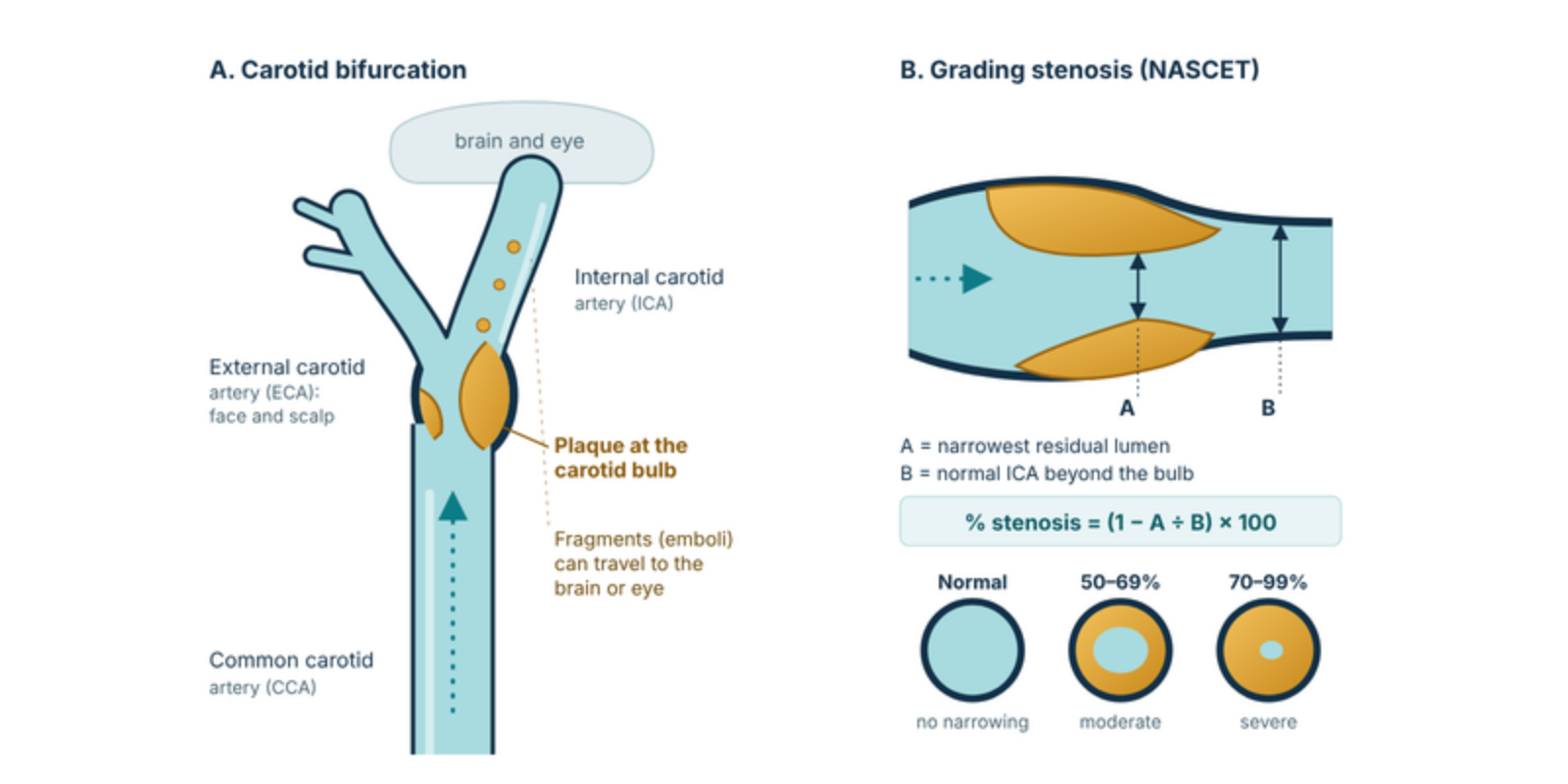
A common carotid artery runs up each side of the neck. Near the angle of the jaw it divides into the external carotid artery, which supplies the face and scalp, and the internal carotid artery (ICA), which supplies the brain and the eye. Atherosclerotic plaque forms most often at the origin of the ICA, in a slight widening called the carotid bulb.
Plaque causes stroke mostly by embolism: fragments of plaque or clot on its surface break away and travel to the brain or eye. Less often, a very tight stenosis or occlusion reduces blood flow to the brain when the collateral circulation is poor. The tightness of the narrowing is only part of the picture. Soft, ulcerated plaque, or plaque with bleeding inside it (intraplaque haemorrhage), sheds more readily than heavily calcified, stable plaque.
The diagram at the top of this page shows where plaque forms and how the degree of narrowing is measured. Treatment thresholds in the major trials and guidelines are based on NASCET-style measurement. The older ECST method compares the lumen with the estimated original width of the bulb, so it gives higher percentages: a 50% NASCET stenosis corresponds to roughly 70% by ECST[25]. When a report gives a percentage, it is worth checking which method was used.
How common is it?
Carotid plaque becomes very common with age, but narrowing severe enough to change management is much less common. In a pooled analysis of four population studies (23,706 people), stenosis of 50% or more without symptoms ranged from 0.2% in men under 50 to 7.5% in men aged 80 and over. Rates in women were lower at every age[3]. A global modelling study estimated that about 1.5% of adults aged 30–79 had a carotid stenosis of 50% or more in 2020, and that prevalence was rising with age and was higher in men[4].
| Group | Stenosis 50% or more | Stenosis 70% or more |
|---|---|---|
| Men under 50 | 0.2% | 0.1% |
| Men 80 and over | 7.5% | 3.1% |
| Women under 50 | 0% | 0% |
| Women 80 and over | 5.0% | 0.9% |
Carotid stenosis is commonly estimated to account for around 10–15% of ischaemic strokes. Other important causes include cardiac embolism (notably atrial fibrillation) and small-vessel disease, which is why a stroke work-up always looks beyond the carotid arteries. Carotid disease also travels with coronary and peripheral artery disease, so a person found to have it should be assessed for atherosclerosis elsewhere.
Causes and risk factors
The great majority of cases are atherosclerosis. The factors that predict moderate or severe stenosis in population data are age, male sex, existing vascular disease, higher blood pressure, an unfavourable cholesterol ratio, diabetes and current smoking[3].
- Not modifiable: increasing age, male sex, family history of premature vascular disease.
- Modifiable: smoking, high blood pressure, high LDL-cholesterol, diabetes, obesity, physical inactivity, chronic kidney disease.
- Other: previous neck radiotherapy (stenosis can appear years later), and known coronary or peripheral artery disease.
Less common causes include carotid dissection, fibromuscular dysplasia, inflammatory arteritis (Takayasu arteritis, giant cell arteritis) and carotid webs. They are more likely in younger patients or those without the usual risk factors, and are managed differently.
Symptoms, examination and natural history
Symptomatic and asymptomatic stenosis
A carotid stenosis is called symptomatic if the patient has had a transient ischaemic attack (TIA), amaurosis fugax, retinal artery occlusion or stroke in the territory of that artery within the last six months[1]. It is asymptomatic if there have been no such events. The distinction drives almost every treatment decision.
- Amaurosis fugax: sudden loss of vision in one eye, often described as a curtain or shade, lasting seconds to minutes and then clearing.
- Hemispheric TIA: sudden weakness, clumsiness or numbness of the opposite arm, leg or face, or difficulty finding or understanding words, resolving within 24 hours (usually within the hour).
- Stroke: the same symptoms persisting beyond 24 hours, or with damage on brain imaging.
Isolated dizziness, light-headedness or blackouts are seldom caused by carotid disease. Most people with a significant stenosis have no symptoms at all. It is found by chance, from a bruit, on imaging done for another reason, or during assessment for other vascular disease.
Examination
A carotid bruit is a weak guide: many people with severe stenosis have none, and many bruits come from other sources. Useful examination includes blood pressure in both arms, pulse rhythm (atrial fibrillation), heart murmurs, peripheral pulses, fundoscopy (cholesterol emboli, called Hollenhorst plaques, in the retinal arteries) and a full neurological examination.
What happens without treatment
Symptomatic stenosis carries a high risk that is heavily front-loaded, greatest in the first days and weeks after the event. In NASCET, patients with a recently symptomatic 70–99% stenosis treated with medical therapy alone had a 26% risk of ipsilateral stroke at two years[5]. That trial used the medical therapy of the 1980s, but the early risk remains the reason for urgency.
Asymptomatic stenosis carries a much lower risk, and it has fallen with modern medical therapy. In the asymptomatic trial ACST-1 (enrolled 1993–2003), the five-year stroke risk without surgery was 11.8%[9]. In the intensive-medical-therapy arms of CREST-2, the four-year risk of the study outcome was 5.3–6.0%[14], after the first 44 days, ipsilateral ischaemic stroke ran at about 1.3–1.7% a year in the medical-therapy-only groups.
Investigations
Carotid duplex ultrasound is the first test. Screening of the general population is not recommended. Testing is directed at people with symptoms, and at selected people who are at high risk and would consider treatment if a severe stenosis were found[2].
| Test | Role and notes |
|---|---|
| Carotid duplex ultrasound | First-line, non-invasive, no radiation. Grades stenosis, shows plaque character and flow. A peak systolic velocity of 125–230 cm/s suggests 50–69% stenosis and above 230 cm/s suggests 70% or more[24]. Results depend on the laboratory, and near-occlusion can be missed. |
| CT angiography (arch to vertex) | Confirms the grade, shows calcification and ulceration, arch anatomy and any intracranial disease. Essential for planning stenting or TCAR. Uses iodinated contrast and radiation. |
| MR angiography | No radiation or iodinated contrast. Can show intraplaque haemorrhage. May overestimate the degree of stenosis. |
| Brain CT or MRI | After TIA or stroke: excludes haemorrhage and defines the infarct. In asymptomatic patients, a silent infarct in the territory of the stenosis marks higher stroke risk. |
| Heart rhythm and echocardiography | ECG and prolonged rhythm monitoring to find atrial fibrillation, which often coexists and may be the true source of emboli. Echocardiography where indicated. |
| Blood tests | Lipid profile, HbA1c, renal function (before contrast), full blood count. |
| Selected tests | Transcranial Doppler (detects spontaneous emboli), cerebrovascular reserve testing and plaque MRI are used in some centres to refine risk in asymptomatic patients. They are not needed for a routine referral. Catheter angiography is now rarely required. |
Medical therapy, and when to operate
Best medical therapy is for every patient
Whatever the decision about a procedure, every patient with carotid stenosis needs intensive treatment of vascular risk. In CREST-2, the intensive medical management protocol set a systolic blood pressure goal below 130 mmHg and an LDL-cholesterol goal below 70 mg/dL (1.8 mmol/L)[14].
| Element | What it involves |
|---|---|
| Antiplatelet | Aspirin 75–100 mg daily, or clopidogrel 75 mg daily if aspirin is not tolerated. After a high-risk TIA or minor stroke, a short course of dual antiplatelet therapy (typically 21 days) is started early under the stroke team’s direction[21,22]. Antiplatelet plans around a procedure are coordinated with the vascular surgeon. |
| Cholesterol | High-intensity statin (for example atorvastatin 40–80 mg or rosuvastatin 20–40 mg). Target LDL-cholesterol below 1.8 mmol/L. Add ezetimibe, then a PCSK9 inhibitor, if not at target. |
| Blood pressure | Systolic below 130 mmHg where tolerated. Avoid aggressive lowering when there is bilateral severe stenosis or occlusion and symptoms that worsen when blood pressure falls. |
| Diabetes | Individualised glycaemic control, using agents with proven cardiovascular benefit where appropriate. |
| Smoking | Complete cessation is the most effective single change. Offer pharmacotherapy and referral to Quitline (13 78 48). |
| Lifestyle | Mediterranean-style diet, regular physical activity, healthy weight, limited alcohol. |
| Anticoagulation and add-on therapy | Anticoagulation for coexisting atrial fibrillation. In selected patients with carotid and other vascular disease, low-dose rivaroxaban (2.5 mg twice daily) plus aspirin may be considered[23]. |
Symptomatic stenosis: operate early when the stenosis is severe
For recently symptomatic patients the evidence is strong and consistent. Pooled data from NASCET and ECST show that surgery is highly beneficial for a 70–99% stenosis, of marginal benefit for 50–69%, of no benefit for 30–49%, and harmful below 30%[7]. Near-occlusion is a special case in which benefit is uncertain.
Timing matters as much as the degree of stenosis. The pooled analysis found that for a stenosis of 50% or more, only five patients needed to be operated on within two weeks of their last event to prevent one stroke over five years, compared with 125 patients operated on more than 12 weeks after the event[8]. Benefit was also greater in men and in patients aged 75 or older. Both the ESVS and SVS guidelines therefore recommend CEA for suitable recently symptomatic patients with a 70–99% stenosis, support it in selected patients with 50–69%, and advise treating within 14 days of the last event[1,2]. For a patient who has had a stroke, revascularisation is generally timed once they are neurologically stable, and after thrombolysis a short delay is advised[1].
| Study | Patients | Main finding |
|---|---|---|
| NASCET 1991[5] | 659 patients, recent TIA or non-disabling stroke, 70–99% stenosis | Ipsilateral stroke at 2 years: 26% with medical therapy, 9% with CEA. |
| NASCET 1998[6] | 50–69% stenosis | Ipsilateral stroke at 5 years: 22.2% with medical therapy, 15.7% with CEA. |
| Pooled ECST and NASCET, 2003[7] | About 6,000 patients | 5-year absolute risk reduction with CEA: 16.0% for 70–99%, 4.6% for 50–69%, no benefit at 30–49%, harm below 30%. |
| Pooled timing analysis, 2004[8] | Stenosis of 50% or more | Number needed to treat to prevent one stroke at 5 years: 5 if operated within 2 weeks of the last event, 125 if after 12 weeks. Greater benefit in men (9 v 36 in women) and age 75 or over (5 v 18 under age 65). |
Asymptomatic stenosis: medical therapy first, procedures for selected patients
The trials that established surgery for asymptomatic stenosis were conducted before modern statins and blood pressure treatment. ACST-1 randomised 3,120 patients with a stenosis of 60% or more. The five-year risk of stroke (including perioperative events) was 6.4% with immediate CEA and 11.8% with deferral, and the benefit persisted at ten years in patients under 75[9,10].
Medical therapy has improved since then, and three recent trials asked whether a procedure still adds benefit. ECST-2 (2-year interim analysis) found no evidence of benefit from adding revascularisation in patients at low or intermediate predicted stroke risk[13]. SPACE-2 was small and underpowered[12]. CREST-2, published in November 2025, is the largest and most contemporary of the three[14].
| Study | Patients | Main finding |
|---|---|---|
| ACST-1 2004 and 2010[9,10] | 3,120 patients, stenosis 60% or more (1993–2003) | Five-year stroke risk 6.4% with immediate CEA v 11.8% with deferral. 30-day stroke or death 3.1%. Benefit at 10 years in patients under 75. |
| ACST-2 2021[11] | 3,625 patients, stenting v CEA | Death or disabling stroke within 30 days about 1% with either. Fatal or disabling non-procedural stroke at 5 years 2.5% with either. Non-disabling procedural strokes were more frequent with stenting (48 v 29). |
| ECST-2 interim 2025[13] | Asymptomatic or low-to-intermediate risk symptomatic, stenosis 50% or more | No evidence of benefit from adding revascularisation to optimised medical therapy in the first 2 years. |
| CREST-2 2025[14] | 2,485 patients with asymptomatic stenosis of 70% or more, in two parallel trials. All received intensive medical management. | Stenting trial: primary outcome 2.8% with stenting v 6.0% with medical therapy alone (P=0.02). Endarterectomy trial: 3.7% v 5.3% (P=0.24). Primary outcome: any stroke or death within 44 days, or ipsilateral ischaemic stroke up to 4 years. |
How should this be read? Intensive medical therapy is now the starting point for every patient with asymptomatic stenosis. In CREST-2, stenting did lower stroke risk, but the difference was about three percentage points over four years, disabling strokes were uncommon in every group, and stenting carried early procedural events (in the first weeks, eight events in the stented group and none in the medical group)[14]. The endarterectomy trial did not show a significant benefit, and the accompanying editorial concluded that there is no longer a role for routine CEA in asymptomatic stenosis[27]. The ESVS and SVS guidelines were written before CREST-2 and are expected to be revised. They differ: SVS recommends CEA for low-risk patients with a stenosis of 70–99%, whereas ESVS advises intervention only when features of higher stroke risk are present[1,2].
Features that ESVS uses to identify a higher-risk asymptomatic stenosis include a silent infarct in the same territory on CT or MRI, progression of the stenosis, echolucent or large plaque, intraplaque haemorrhage on MRI, spontaneous emboli on transcranial Doppler and impaired cerebrovascular reserve. Even then, ESVS advises a procedure only when life expectancy exceeds five years and the operator’s 30-day stroke and death rate is 3% or lower[1]. For a patient with an asymptomatic stenosis, the sensible starting point is intensive medical therapy at once, with a procedure discussed only when there is a specific reason to think this patient is at higher risk than the trials suggest.
Carotid endarterectomy, carotid stenting and TCAR
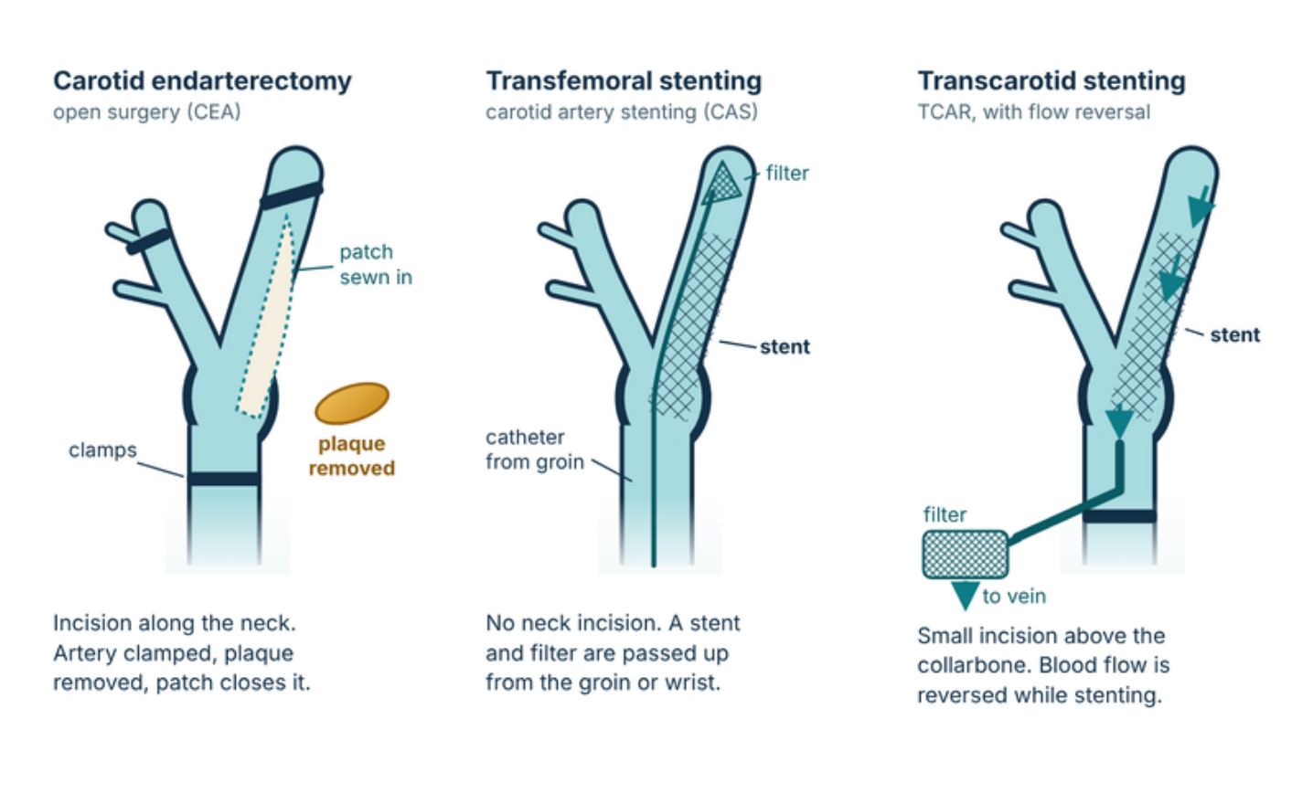
When revascularisation is chosen, there are three options. All three aim to remove or cover the plaque and restore a smooth lumen. They differ in access, in how the brain is protected during the procedure, and in the evidence behind them.
Carotid endarterectomy (CEA)
How it is done. Under general or regional anaesthesia (the GALA trial found no difference in outcome between the two[17]), an incision is made along the front of the neck. The common, internal and external carotid arteries are clamped, sometimes with a temporary shunt to maintain flow to the brain. The artery is opened, the plaque is removed and the artery is closed with a patch, which is preferred to direct closure[1]. Most patients stay one to two nights and are back to normal activity within a couple of weeks.
Who it suits. CEA is the reference standard. It is the preferred option for recently symptomatic patients who need early treatment, for patients aged 70 or over, and for most standard-risk patients.
Evidence. CEA is the procedure tested in NASCET, ECST and ACST-1. In CREST, 30-day stroke was 2.3% with CEA against 4.1% with stenting[15]. ESVS asks that surgeons keep 30-day stroke and death below 6% for symptomatic and 3% for asymptomatic patients[1].
Risks. Stroke, heart attack and death (see Table 6). Injury to a cranial nerve, causing hoarseness, a weak corner of the mouth or tongue deviation, occurred in about 5% in CREST, and most cases recover. Other risks are bleeding in the neck that needs a return to theatre, wound problems, and, uncommonly, hyperperfusion syndrome (headache or seizure from a sudden rise in blood flow to the brain). Restenosis can occur and is usually silent.
Transfemoral carotid stenting (CAS)
How it is done. Through a needle puncture in the groin (or wrist), a catheter is guided through the aorta into the common carotid artery. A filter or balloon is placed beyond the narrowing to catch debris, the artery is dilated and a stent is deployed. It is usually done under local anaesthesia. Dual antiplatelet therapy starts before the procedure and continues for at least a month afterwards.
Who it suits. CAS is considered when the neck is hostile (previous radiotherapy or neck surgery), the bifurcation is high, or there is restenosis after CEA. In CREST, stenting performed relatively better in patients under about 70 and CEA in older patients[15]. It is less suitable for a tortuous or heavily calcified arch, or for very early treatment after symptoms.
Evidence. CREST found similar overall outcomes (7.2% with stenting v 6.8% with CEA for stroke, heart attack, death or later ipsilateral stroke)[15]. Stroke was more common with stenting (4.1% v 2.3%) and heart attack less common (1.1% v 2.3%). In ICSS, stroke, death or procedural heart attack within 120 days in symptomatic patients was 8.5% with stenting v 5.2% with CEA[16]. ESVS recommends CEA over transfemoral stenting when treatment is done in the first 7–14 days after symptoms[1]. In asymptomatic patients, ACST-2 found the two procedures broadly equivalent[11].
Risks. Stroke (higher than CEA in symptomatic trials, especially in older patients and soon after symptoms), a slow heart rate and low blood pressure during the procedure, groin bleeding, contrast-related kidney injury, hyperperfusion syndrome and restenosis of the stent. Cranial nerve injury is rare (0.3% in CREST).
Transcarotid artery revascularisation (TCAR)
How it is done. TCAR is a hybrid of surgery and stenting. Through a small incision just above the collarbone, a sheath is placed directly into the common carotid artery, which avoids passing a catheter through the aortic arch. A second small sheath goes into the femoral vein. The carotid is then clamped below the sheath and blood is diverted so that it runs backwards from the brain, through a filter and back into the vein while the stent is placed. Any debris travels away from the brain rather than towards it. Reversal usually lasts only a few minutes. Antiplatelet therapy is used before and for at least a month after.
Who it suits. TCAR is aimed at patients for whom a stent is preferred to open surgery, such as those with a hostile neck, a high bifurcation or significant cardiac risk, and whose common carotid artery has enough length and calibre on CT angiography. It is not well suited to the first days after symptoms.
Evidence. There are no randomised trials. The single-arm ROADSTER 1 and 2 studies reported 30-day stroke of 1.4% and 1.9%, and stroke or death of 2.8% and 2.3% (intention-to-treat figures)[18,19]. ROADSTER 2 enrolled 692 patients at 43 sites, all at high risk for CEA, and 26% had symptoms[19]. Registry data suggest lower stroke than transfemoral stenting and outcomes broadly similar to CEA overall, with fewer cranial nerve injuries. When TCAR was done within 14 days of neurological symptoms, however, a matched registry comparison found more ipsilateral strokes than with CEA (3.8% v 1.8%)[20], and ESVS notes higher in-hospital stroke when TCAR is done within two days of symptoms (5.6%, against 2.5% at 3–14 days)[1]. SVS guidelines favour TCAR over transfemoral stenting, and over CEA in high surgical risk patients, while ESVS is more cautious because of the lack of randomised data[1,2].
Availability in Australia. At the time of writing, TCAR in Australia is available through the TGA Special Access pathway at a small number of centres[26]. Availability in Western Australia should be confirmed when a referral is made.
Risks. Stroke, injury at the neck access site (bleeding, dissection), cranial nerve injury (less frequent than after CEA but not absent), rarely intolerance of flow reversal, a slow heart rate or low blood pressure, heart attack and restenosis.
| Feature | Carotid endarterectomy | Transfemoral stenting | TCAR |
|---|---|---|---|
| Access | Neck incision; artery opened | Needle puncture in groin or wrist; catheter through the aortic arch | Small incision above the collarbone; sheath directly in the carotid |
| Brain protection | Clamping, with selective shunt | Filter or balloon beyond the lesion | Flow reversal through a filter |
| Anaesthesia | General or regional (equivalent outcomes)[17] | Local, with or without sedation | Local or general |
| Antiplatelets | Aspirin, continued through surgery | Dual therapy before and for at least a month after | Dual therapy before and for at least a month after |
| Randomised evidence | Extensive (NASCET, ECST, ACST-1, ACST-2, CREST, ICSS, CREST-2) | Extensive (CREST, ICSS, ACST-2, CREST-2) | None. Single-arm trials and registry data only |
| 30-day stroke | 2.3% in CREST[15]. Death or disabling stroke about 1% in ACST-2 (asymptomatic)[11] | 4.1% in CREST[15]. Death or disabling stroke about 1% in ACST-2 (asymptomatic)[11] | 1.4–1.9% in ROADSTER 1 and 2 (high-risk patients)[18,19]. Higher than CEA if done within 14 days of symptoms[20] |
| 30-day heart attack | 2.3% in CREST[15] | 1.1% in CREST[15] | 0.9% in ROADSTER 2[19] |
| Cranial nerve injury | About 5% in CREST, mostly temporary | Rare (0.3% in CREST) | Lower than CEA, not zero |
| Best suited to | Most standard-risk patients; recently symptomatic patients needing early treatment; age 70 or over | Hostile neck, high bifurcation, restenosis after CEA; younger patients; patient preference in asymptomatic disease | Where a stent is preferred and the common carotid anatomy is suitable |
| Less suited to | Hostile neck, high bifurcation | Early after symptoms; age 70 or over; tortuous or calcified arch | Within 2 weeks of symptoms; short or small common carotid |
| Availability | Widely available | Selected centres | Limited in Australia; confirm at referral |
Choosing between procedures
The choice is individual. It rests on the timing and nature of symptoms, the degree of stenosis, the anatomy on CT angiography, age, cardiac and other risk, and the audited results of the operator. As a working rule, CEA is the first choice for recently symptomatic patients and for most standard-risk patients. Stenting, by transfemoral or transcarotid access, is considered for patients with a hostile neck or high bifurcation, for restenosis, and for selected patients where an open operation carries more risk than a stent. Since CREST-2, stenting is also a reasonable option for the small group of asymptomatic patients in whom a procedure is chosen. The patient’s own preferences matter, and the decision should be shared.
For referring doctors
The flow chart below sets out how to triage a patient with suspected or confirmed carotid stenosis. The two branches are urgent when the patient has recent symptoms, and routine when they do not. It reflects the evidence above and the ESVS and SVS guidelines, and it is a guide only. Please phone to discuss any case that does not fit. Our general GP referral guidelines cover other vascular conditions.
- Decision point
- Urgent
- Vascular surgical opinion
- Medical management
Suspected or confirmed carotid stenosisBruit, vascular disease, neurological or visual event, or a finding on imaging
Any neurological or retinal symptoms in the last 6 months?TIA, amaurosis fugax, retinal artery occlusion, or stroke in the carotid territory
Same-day assessmentEmergency department or a rapid-access TIA and stroke service. Brain imaging to exclude haemorrhage. Antiplatelet and statin per the stroke team.
Urgent carotid imagingDuplex, then CT or MR angiography of the head and neck. Ask for the stenosis to be reported by the NASCET method.
Stenosis of the artery on the symptomatic side?
70–99%: urgent vascular surgical referralAim to revascularise within 14 days of the last event. CEA is the first choice.
50–69%: vascular surgical opinionRevascularisation is reasonable in selected patients, particularly men and those with a recent event. Decide with the vascular and stroke teams.
Under 50%, or occlusion: medical therapyLook for another source (cardiac, small vessel). If near-occlusion, seek an opinion.
Carotid duplex and start best medical therapy nowAntiplatelet, statin, blood pressure control, stop smoking. No urgency, and no procedure is needed for the diagnosis alone.
Degree of stenosis (NASCET)?
Under 50%Risk factor control. No intervention.
50–59%Best medical therapy and periodic surveillance duplex.
60–99%: routine vascular surgical referralBest medical therapy is the default. A procedure is considered only for selected patients: features of higher stroke risk (for example silent infarct on imaging, progression, echolucent plaque), life expectancy over 5 years, and a low procedural risk.
For every patientAntiplatelet, statin (LDL-cholesterol below 1.8 mmol/L), systolic blood pressure below 130 mmHg where tolerated, smoking cessation, glycaemic control, exercise and diet. Teach the warning signs of stroke and to call 000.
What to include in a referral
- The date and description of any neurological or visual event, and which side it affected.
- Carotid duplex, CT or MR angiography and brain imaging: the reports, and the images where possible. Please state whether the percentage stenosis is by NASCET or ECST.
- Current medicines, especially antiplatelets and anticoagulants, and any contrast allergy.
- Renal function, HbA1c and lipid results.
- Relevant history: cardiac disease, atrial fibrillation, previous neck surgery or radiotherapy, diabetes, and general fitness.
While the patient waits
- Start or continue antiplatelet therapy and a high-intensity statin. Do not stop antiplatelet therapy for a planned procedure unless advised by the surgeon.
- Treat blood pressure, but avoid abrupt lowering in symptomatic patients with bilateral severe stenosis or occlusion.
- Give clear advice about warning symptoms and driving, in line with the Austroads guidance on fitness to drive.
Contact Perth Vascular Clinic
For an urgent case, please phone to discuss it with us on 08 6116 4955. A patient with a suspected TIA or stroke should go to an emergency department first.
Suite 218, St John of God Medical Clinic, 25 McCourt Street, Subiaco WA 6008.
Request an AppointmentCall 08 6116 4955After treatment
Best medical therapy continues for life, whether or not a procedure was done. After CEA, stenting or TCAR, duplex ultrasound is used to check the treated artery for restenosis and to follow the other carotid artery. Follow-up is commonly at about a month and then at intervals set by the surgeon. Patients should know the warning signs of stroke and to call 000. Our discharge information for patients after carotid endarterectomy and after carotid stenting sets out what to expect in the first weeks at home.
References
- Naylor R, Rantner B, Ancetti S, et al. European Society for Vascular Surgery (ESVS) 2023 clinical practice guidelines on the management of atherosclerotic carotid and vertebral artery disease. Eur J Vasc Endovasc Surg 2023;65:7–111.
- AbuRahma AF, Avgerinos ED, Chang RW, et al. Society for Vascular Surgery clinical practice guidelines for management of extracranial cerebrovascular disease. J Vasc Surg 2022;75:4S–22S.
- de Weerd M, Greving JP, Hedblad B, et al. Prevalence of asymptomatic carotid artery stenosis in the general population: an individual participant data meta-analysis. Stroke 2010;41:1294–1297.
- Song P, Fang Z, Wang H, et al. Global and regional prevalence, burden, and risk factors for carotid atherosclerosis: a systematic review, meta-analysis, and modelling study. Lancet Glob Health 2020;8:e721–e729.
- North American Symptomatic Carotid Endarterectomy Trial Collaborators. Beneficial effect of carotid endarterectomy in symptomatic patients with high-grade carotid stenosis. N Engl J Med 1991;325:445–453.
- Barnett HJM, Taylor DW, Eliasziw M, et al. Benefit of carotid endarterectomy in patients with symptomatic moderate or severe stenosis. N Engl J Med 1998;339:1415–1425.
- Rothwell PM, Eliasziw M, Gutnikov SA, et al. Analysis of pooled data from the randomised controlled trials of endarterectomy for symptomatic carotid stenosis. Lancet 2003;361:107–116.
- Rothwell PM, Eliasziw M, Gutnikov SA, Warlow CP, Barnett HJM. Endarterectomy for symptomatic carotid stenosis in relation to clinical subgroups and timing of surgery. Lancet 2004;363:915–924.
- Halliday A, Mansfield A, Marro J, et al. Prevention of disabling and fatal strokes by successful carotid endarterectomy in patients without recent neurological symptoms: randomised controlled trial (ACST-1). Lancet 2004;363:1491–1502.
- Halliday A, Harrison M, Hayter E, et al. 10-year stroke prevention after successful carotid endarterectomy for asymptomatic stenosis (ACST-1): a multicentre randomised trial. Lancet 2010;376:1074–1084.
- Halliday A, Bulbulia R, Bonati LH, et al. Second asymptomatic carotid surgery trial (ACST-2): a randomised comparison of carotid artery stenting versus carotid endarterectomy. Lancet 2021;398:1065–1073.
- Reiff T, Eckstein HH, Mansmann U, et al. Carotid endarterectomy or stenting or best medical treatment alone for moderate-to-severe asymptomatic carotid artery stenosis: 5-year results of SPACE-2. Lancet Neurol 2022;21:877–888.
- Donners SJA, van Velzen TJ, Cheng SF, et al. Optimised medical therapy alone versus optimised medical therapy plus revascularisation for asymptomatic or low-to-intermediate risk symptomatic carotid stenosis (ECST-2): 2-year interim results of a multicentre randomised trial. Lancet Neurol 2025;24:389–399.
- Brott TG, Howard G, Lal BK, et al. Medical management and revascularization for asymptomatic carotid stenosis (CREST-2). N Engl J Med 2025; published 21 November 2025. doi:10.1056/NEJMoa2508800.
- Brott TG, Hobson RW 2nd, Howard G, et al. Stenting versus endarterectomy for treatment of carotid-artery stenosis (CREST). N Engl J Med 2010;363:11–23.
- International Carotid Stenting Study investigators. Carotid artery stenting compared with endarterectomy in patients with symptomatic carotid stenosis (ICSS): an interim analysis of a randomised controlled trial. Lancet 2010;375:985–997.
- GALA Trial Collaborative Group. General anaesthesia versus local anaesthesia for carotid surgery (GALA): a multicentre, randomised controlled trial. Lancet 2008;372:2132–2142.
- Kwolek CJ, Jaff MR, Leal JI, et al. Results of the ROADSTER multicenter trial of transcarotid stenting with dynamic flow reversal. J Vasc Surg 2015;62:1227–1234.
- Kashyap VS, Schneider PA, Foteh M, et al. Early outcomes in the ROADSTER 2 study of transcarotid artery revascularization in patients with significant carotid artery disease. Stroke 2020;51:2620–2629.
- Elmously A, Rich N, Lazar AN, et al. Outcomes of early transcarotid artery revascularization versus carotid endarterectomy after acute neurologic events. J Vasc Surg 2022;76:760–768.
- Wang Y, Wang Y, Zhao X, et al. Clopidogrel with aspirin in acute minor stroke or transient ischemic attack (CHANCE). N Engl J Med 2013;369:11–19.
- Johnston SC, Easton JD, Farrant M, et al. Clopidogrel and aspirin in acute ischemic stroke and high-risk TIA (POINT). N Engl J Med 2018;379:215–225.
- Eikelboom JW, Connolly SJ, Bosch J, et al. Rivaroxaban with or without aspirin in stable cardiovascular disease (COMPASS). N Engl J Med 2017;377:1319–1330.
- Grant EG, Benson CB, Moneta GL, et al. Carotid artery stenosis: gray-scale and Doppler US diagnosis. Society of Radiologists in Ultrasound Consensus Conference. Radiology 2003;229:340–346.
- Rothwell PM, Gibson RJ, Slattery J, Sellar RJ, Warlow CP. Equivalence of measurements of carotid stenosis: a comparison of three methods on 1001 angiograms. Stroke 1994;25:2435–2439.
- Gosford Hospital leading Australia with new lifesaving stroke prevention surgery from US. ABC News, 15 April 2026.
- Brown MM, Bonati LH. Editorial accompanying the CREST-2 report. N Engl J Med 2025.
General information only. This article is written for patients and referring doctors and is not a substitute for individual medical advice. Treatment decisions depend on your symptoms, imaging, general health and preferences, and should be made with your doctor and a vascular surgeon. Risk figures come from clinical trials and registries and describe groups of patients, not any one person.

Listen to the Latest Podcast
Listen to the latest podcast featuring Dr Marek Garbowski and Medical Forum, WA’s independent monthly publication for health professionals.
Get in Touch

About the Author: Dr Marek Garbowski
Dr Marek Garbowski, MBBS, FRACS (Vasc), is a Vascular and Endovascular Surgeon and the founder of Perth Vascular Clinic, which he established in Subiaco in 2006.
He completed his MBBS in 1994 and trained across Australia and New Zealand before being awarded Fellowship of the Royal Australasian College of Surgeons in Vascular Surgery in 2005. He performs both open vascular surgery and minimally invasive endovascular procedures, with particular interests in carotid stenting, complex aortic aneurysm repair, endovenous treatment of varicose veins and the management of leg and foot ulcers.
Dr Garbowski consults in Subiaco and Joondalup, heads the Vascular and Endovascular Surgery Department at Joondalup Health Campus, and teaches at UWA and Notre Dame.

Millions of Women suffer from Pelvic Pain
In some cases, this pain can be caused by Pelvic Venous Disorders (also known as Pelvic Congestion Syndrome).
If you're in pain and can't seem to find any answers, read our guide and find out more.