
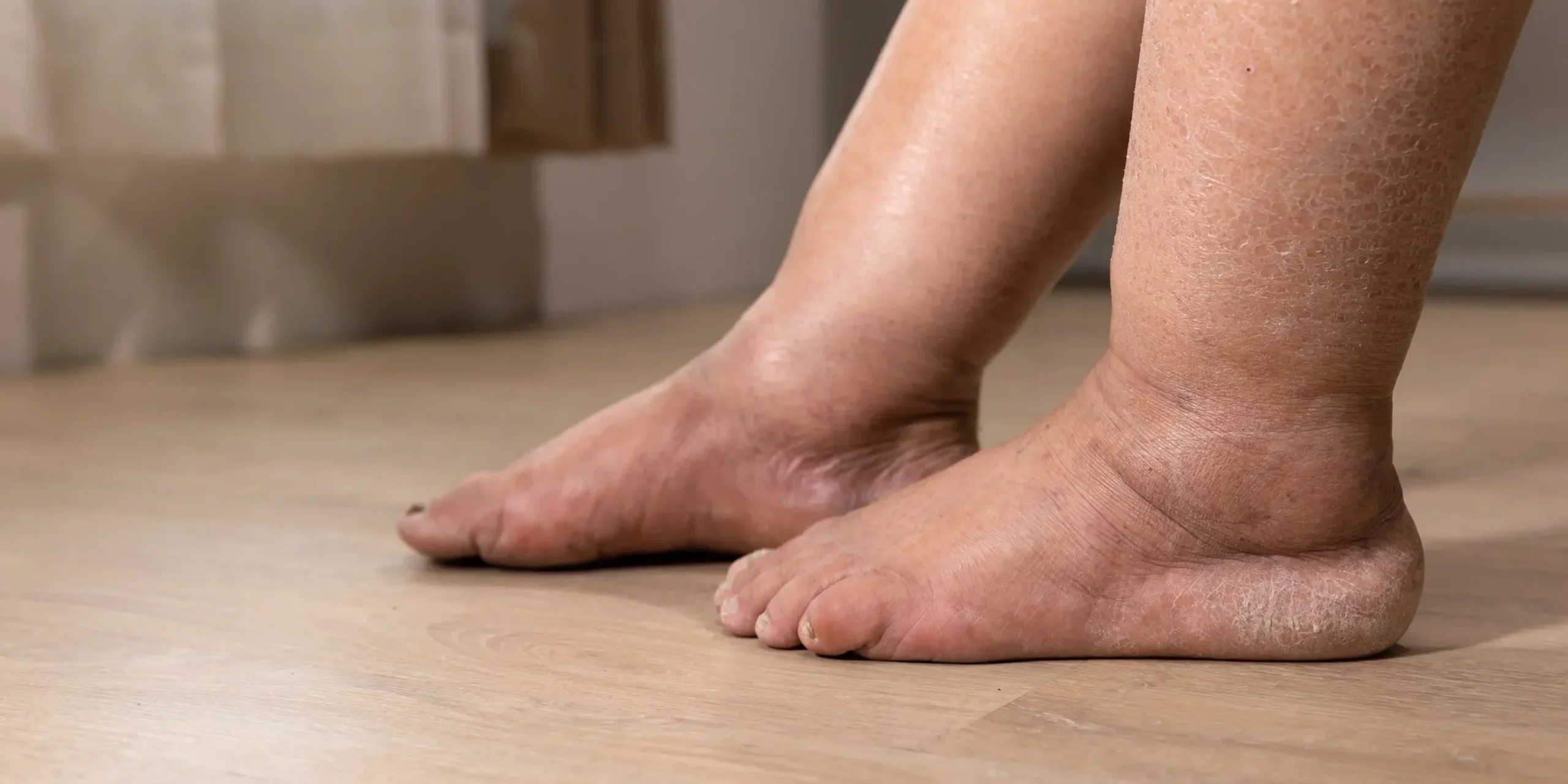
“Swollen legs” is a symptom, not a diagnosis. Three quite different problems account for most large or heavy legs – oedema, lymphoedema and lipoedema – and each needs a different plan. A guide for patients and general practitioners.
In this article
At a glance
- Oedema is excess fluid in the tissues. It is usually caused by something upstream – the veins, the heart, kidneys, liver, a medication, or simply prolonged sitting and standing. In otherwise well adults the most common cause is chronic venous insufficiency. It pits when pressed and often improves overnight.
- Lymphoedema is swelling from a lymphatic drainage system that is not coping. It is chronic, protein-rich, tends to involve the foot and toes, and gradually hardens the tissues.
- Lipoedema is a chronic disorder of fat (adipose tissue), almost always in women. Fat builds up symmetrically on both legs – often hips to ankles – with pain, tenderness and easy bruising, while the feet are spared. It does not respond to diet like ordinary weight gain.
- The three can overlap. Venous disease can cause oedema and eventually lymphoedema (“phlebolymphoedema”); obesity can coexist with lipoedema and, when severe, overload the lymphatics. A careful history and examination, with targeted tests, almost always sorts them out.
For GPs: the questions that discriminate best are – one leg or both? Are the feet involved? Does it pit? Is it painful to touch? Does it go down overnight? And has it changed with weight loss?
For patients: if your legs are swollen, uncomfortable or changing shape, see your GP. Most causes are treatable, and early diagnosis prevents progression.
How the leg normally handles fluid and fat
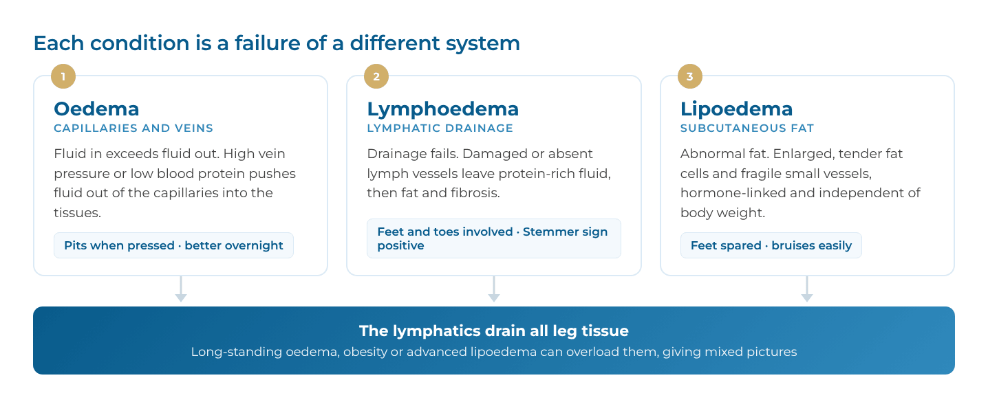
Leg volume depends on a balance between three systems, and each condition is a failure of a different one.
- Capillaries and veins. Blood pressure pushes fluid out of the capillaries into the tissues every minute. The veins carry blood back to the heart, helped by one-way valves and the calf-muscle pump. If venous pressure rises (valve failure, obstruction, heart failure) or the blood’s protein level falls (kidney or liver disease, malnutrition), more fluid leaks out than can be removed – this is oedema.
- Lymphatics. Several litres of fluid and protein filter into the tissues each day, and the lymphatic vessels carry them back through lymph nodes to the bloodstream. Fluid that is not reabsorbed is cleared by the lymphatics, so they are the safety valve. If they are damaged, absent or overwhelmed, protein-rich fluid accumulates, triggering inflammation, fat deposition and fibrosis – this is lymphoedema.
- Subcutaneous fat. The fat layer under the skin normally stores energy and changes in proportion to overall body weight. In lipoedema, the fat in the legs (and sometimes arms) behaves abnormally – enlarged fat cells, fragile small vessels and low-grade inflammation – independent of total body weight.
Because the lymphatics drain all leg tissues, long-standing oedema of any cause, severe obesity and advanced lipoedema can all eventually overload them. That is why mixed pictures are common in older or heavier patients.
Lower leg oedema
Oedema is the most common of the three and is a sign of another process rather than a disease in itself. Its pattern – one leg or both, sudden or gradual – points to the cause.
Common causes
| Pattern | Likely causes |
|---|---|
| Both legs, gradual | Chronic venous insufficiency; heart failure; kidney disease (nephrotic syndrome, CKD); liver cirrhosis; low albumin (malnutrition, protein-losing states); pulmonary hypertension and obstructive sleep apnoea; hypothyroidism; pregnancy; prolonged sitting (“dependent” or “armchair” oedema) |
| Both legs, medication-related | Calcium-channel blockers (especially amlodipine, dose-related); gabapentin and pregabalin; NSAIDs; corticosteroids; oestrogens and some hormonal therapies; glitazones; minoxidil |
| One leg, acute (days) | Deep vein thrombosis until proven otherwise; acute superficial thrombophlebitis; cellulitis; ruptured Baker’s cyst; muscle injury or haematoma |
| One leg, gradual (weeks to months) | Venous insufficiency or post-thrombotic syndrome; iliac vein compression (May-Thurner, usually the left leg); pelvic or groin mass compressing veins or lymphatics; early lymphoedema |
Chronic venous insufficiency: the leading cause
Chronic venous insufficiency (CVI) is the single most common cause of chronic leg swelling in otherwise well adults. It is often missed when varicose veins are small, hidden by body habitus, or absent – swelling can be the first and only sign.
How it develops. Normally, one-way valves and the calf-muscle pump empty the leg veins with each step. In CVI this fails, leaving the veins under sustained high pressure whenever the person is upright (ambulatory venous hypertension). Fluid leaks into the tissues, low-grade inflammation follows, and over years the skin is damaged. The three mechanisms are:
- Reflux: valve failure in the superficial (saphenous), perforator or deep veins; usually primary and often familial
- Obstruction: after deep vein thrombosis (post-thrombotic syndrome) or from iliac vein compression (May-Thurner)
- Pump failure: immobility, a stiff ankle, obesity or weak calf muscles
Risk factors: increasing age, female sex and pregnancy, family history, occupations with prolonged standing or sitting, obesity, previous DVT or leg injury.
Severity is graded with the international CEAP clinical classification:
| CEAP class | Clinical findings |
|---|---|
| C0 | No visible signs (symptoms only) |
| C1 | Spider (telangiectatic) or reticular veins |
| C2 | Varicose veins |
| C3 | Venous oedema |
| C4 | Skin changes – pigmentation or eczema (C4a); lipodermatosclerosis or atrophie blanche (C4b); corona phlebectatica at the ankle (C4c) |
| C5 | Healed venous ulcer |
| C6 | Active venous ulcer |
A venous duplex ultrasound shows exactly which veins reflux or are obstructed, and therefore which treatment will help. Correcting the venous problem treats the swelling at its source rather than just containing it.
Acute superficial thrombophlebitis
Superficial thrombophlebitis (superficial vein thrombosis) is a clot in a vein just under the skin, usually a varicose vein. It causes a hot, red, tender, cord-like vein, often with swelling of the surrounding leg. It is not a trivial condition: around one in four patients has a coexisting DVT at presentation, and clot close to the groin (saphenofemoral) or knee (saphenopopliteal) junction can extend into the deep veins and cause pulmonary embolism.
- Risk factors include varicose veins, recent surgery or immobility, pregnancy and oestrogen therapy, cancer, thrombophilia and intravenous cannulas
- It needs prompt assessment with a venous duplex – the same or next day – to measure the clot’s length and distance from the deep veins
- Treatment is guided by the scan: compression and anti-inflammatories for short segments away from the junctions; a course of prophylactic-dose anticoagulation (e.g. fondaparinux for about 45 days) for longer segments; clot within about 3 cm of a junction is treated as a DVT
- Recurrent episodes, or thrombophlebitis in non-varicose veins, warrant a search for an underlying cause such as cancer or a clotting disorder
- Once settled, treating the underlying varicose veins reduces the chance of recurrence
Typical symptoms and signs
- Swelling that is worse at the end of the day and after standing, and better overnight or with elevation
- Ankle and shin swelling that pits – a thumb pressed for 10 seconds leaves a dent
- Aching, heaviness and tight shoes or sock marks
- With venous disease: varicose veins, ankle flare, brown pigmentation, eczema, hardened skin (lipodermatosclerosis) and eventually ulceration
- With systemic causes: breathlessness, orthopnoea, weight gain, frothy urine, jaundice or fatigue may give the clue
How common is it?
Leg swelling becomes more common with age. Chronic venous disease is the most frequent single cause in otherwise well adults, and visible varicose veins are present in roughly a quarter to a third of adults in population studies. Drug-induced oedema is common and often missed; it does not respond well to diuretics and usually settles when the drug is reduced or changed.
Lymphoedema
Lymphoedema is chronic swelling caused by reduced lymphatic transport. Unlike simple oedema, the trapped fluid is rich in protein, which drives inflammation, fat deposition and progressive fibrosis – so untreated lymphoedema slowly becomes firmer, larger and harder to reverse.
Causes
Secondary lymphoedema (the majority) follows damage to a previously normal lymphatic system:
- Cancer treatment – lymph node dissection in the groin or pelvis (melanoma, gynaecological, vulval, prostate and bladder cancers) and radiotherapy
- Pelvic or groin tumours, including recurrence – new unilateral leg lymphoedema in an adult needs this excluded
- Trauma, extensive surgery or scarring around the knee or groin; vein harvesting
- Recurrent cellulitis, which damages lymphatics and is also a consequence of lymphoedema
- Long-standing venous disease (phlebolymphoedema) and severe obesity (obesity-related lymphoedema)
- Filariasis – the leading cause worldwide, but rarely seen in Australia outside returned travellers and migrants
Primary lymphoedema results from a developmental abnormality of the lymphatics, sometimes genetic and familial. It may appear at birth (e.g. Milroy disease), around puberty or in early adulthood (the most common pattern), or later in life.
Stages (International Society of Lymphology 2023)
| ISL stage | What it looks like |
|---|---|
| 0 – latent | Lymphatic transport is impaired but there is no visible swelling yet; may cause heaviness. Can last months or years |
| I – early | Soft swelling that pits and largely subsides with elevation |
| II – established | Elevation alone rarely reduces it; pitting is obvious early in this stage but lessens as fat and fibrosis develop |
| III – lymphostatic elephantiasis | Large, firm limb without pitting; skin thickening, deep folds, warty growths (papillomatosis), lymph leakage |
Typical symptoms and signs
- Swelling usually starts distally, involving the foot and toes (“squared-off” toes, a dorsal foot “buffalo hump”)
- Often one leg, or both but asymmetric
- Positive Stemmer sign – the skin at the base of the second toe cannot be pinched and lifted
- Heaviness and tightness rather than tenderness; pain is not a dominant feature
- Over time: skin thickening, deepened skin creases, hyperkeratosis, papillomatosis and lymphorrhoea (weeping)
- Repeated episodes of cellulitis – a red, hot, painful limb, often with fever
Lipoedema
Lipoedema is a chronic disease of the subcutaneous fat, not a fluid problem and not simply obesity. In January 2026 the Lipedema World Alliance published the first international Delphi consensus (71 experts and patient representatives, 19 countries, 59 agreed statements), which now forms the best reference definition. The key points below follow that consensus.
What it is
- Symmetrical, bilateral enlargement of fat in the limbs – out of proportion to the trunk – accompanied by pain or discomfort
- The feet (and hands) are typically spared, often with a visible “cuff” or step at the ankle
- The arms are involved in a substantial proportion of patients (reported 30-80%)
- Affected tissue is tender to touch or pressure, feels heavy, and bruises easily
- Pitting oedema is usually absent, and the Stemmer sign is usually negative – unless lymphatic overload has developed
Who gets it
- Almost exclusively women; in men it is rare and usually linked to hormonal abnormalities
- Onset or worsening typically follows hormonal change – puberty, pregnancy or menopause
- A family history is common, suggesting a hereditary component
- True prevalence is unknown; estimates range from under 1% to about 12% of adult women
- It occurs at any body weight. Obesity frequently coexists but is a separate disease – lipoedema is not caused by obesity, and lipoedema fat is largely resistant to diet and persists even after bariatric surgery
Why it happens
The cause is not fully understood. Current evidence points to abnormal subcutaneous fat (enlarged fat cells), low-grade inflammation, hormonal (oestrogen) influences, fragile and leaky small vessels, and increased extracellular fluid bound within the tissue. The lymphatic system is usually normal early on, but can become overloaded later.
Stages and types
Traditional clinical stages describe the skin and fat texture:
| Stage | Typical appearance |
|---|---|
| I | Smooth skin; soft, evenly enlarged fat with small palpable nodules |
| II | Uneven, dimpled skin surface; larger nodules |
| III | Large lobules and overhanging folds, especially at the inner knee and thighs; deformity |
| Lipo-lymphoedema (sometimes called stage IV) | Lipoedema with secondary lymphoedema – foot swelling and a positive Stemmer sign appear |
The distribution is also described by type (buttocks and hips; hips to knees; hips to ankles; arms; lower legs only). The 2026 consensus stresses that stage and type are descriptive only: they do not reflect pain or disability and have limited value in guiding treatment, and progression is not inevitable. Weight gain and hormonal events are the main drivers of worsening.
Impact
Lipoedema commonly causes reduced mobility, knee and hip strain (including osteoarthritis), and a substantial psychological burden – body-image distress, anxiety and depression. Many women describe years of being told to “just lose weight”. Missed or delayed diagnosis worsens symptoms, mental wellbeing and costs; recognising the condition is itself therapeutic.
Side by side
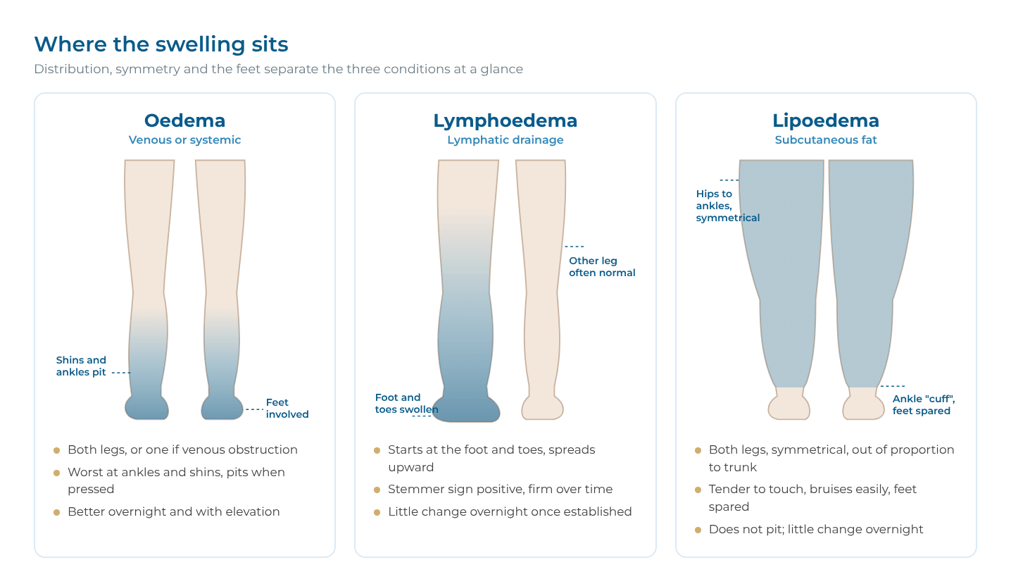
The feet, the pitting test, tenderness and the response to elevation separate the three conditions in most patients.
| Feature | Oedema (venous / systemic) | Lymphoedema | Lipoedema |
|---|---|---|---|
| What accumulates | Low-protein fluid | Protein-rich fluid, then fat and fibrosis | Abnormal fat (± bound fluid) |
| Sex | Both | Both (primary forms more often female) | Almost always women |
| Onset | Any age; often older | Any age; after cancer treatment, trauma or infection | Puberty, pregnancy or menopause |
| Distribution | Ankles and shins; one or both legs | Often one leg or asymmetric; starts distally | Both legs symmetrically, hips/thighs to ankles; ± arms |
| Feet | Involved | Involved – toes and dorsum | Spared; ankle “cuff” |
| Pitting | Yes | Early yes; lost with fibrosis | Usually no |
| Stemmer sign | Negative | Positive | Negative (unless lymphatic overload) |
| Pain / tenderness | Ache, heaviness | Heaviness; little tenderness | Tender to touch; aching; marked |
| Bruising | No | No | Easy bruising |
| Overnight / elevation | Improves | Improves early only | Little change |
| Skin | Venous pigmentation, eczema, ulcers (if venous) | Thickening, papillomatosis, weeping | Soft, cool; nodular or dimpled |
| Cellulitis | Occasional | Recurrent, common | Uncommon unless lymphoedema develops |
| Response to weight loss | Variable | Helps if obesity contributes | Little effect on the affected fat |
| Diuretics help? | Only for systemic fluid overload | No | No |
Examination: what to look for
Examine both legs standing and lying, with shoes and socks off, from the waist down. Five bedside tests do most of the work.
- Distribution and symmetry. Compare both legs, hips, buttocks and arms. Disproportion between a slim upper body and heavy legs suggests lipoedema; a single swollen limb points to venous obstruction, DVT or lymphoedema.
- The feet. Look at the dorsum and toes. Foot and toe involvement favours lymphoedema or oedema; a clear step at the ankle with a slim foot favours lipoedema.
- Pitting. Press firmly over the shin or ankle for 10 seconds. Deep, easy pitting = oedema or early lymphoedema; little or none = lipoedema or fibrotic lymphoedema.
- Stemmer sign. Try to pinch and lift a fold of skin at the base of the second toe. Inability to do so (positive) indicates lymphoedema.
- Palpation for tenderness and texture. Gentle pinch of the thigh or shin fat: tenderness and small nodules (“rice grains” or “beans”) suggest lipoedema.
Also check:
- Veins and skin: varicose veins, corona phlebectatica at the ankle, pigmentation, eczema, lipodermatosclerosis, ulcer scars
- Pulses and ABPI: essential before any compression therapy; peripheral arterial disease changes the plan
- Lymph node areas: groin and abdomen for masses or surgical scars
- General signs: raised JVP, crackles, ascites, jaundice, thyroid signs, BMI, waist-to-height ratio (more useful than BMI when lipoedema distorts body shape)
- Joint hypermobility: reported more often in lipoedema
- Signs of infection: warmth, spreading redness, fever
Investigations
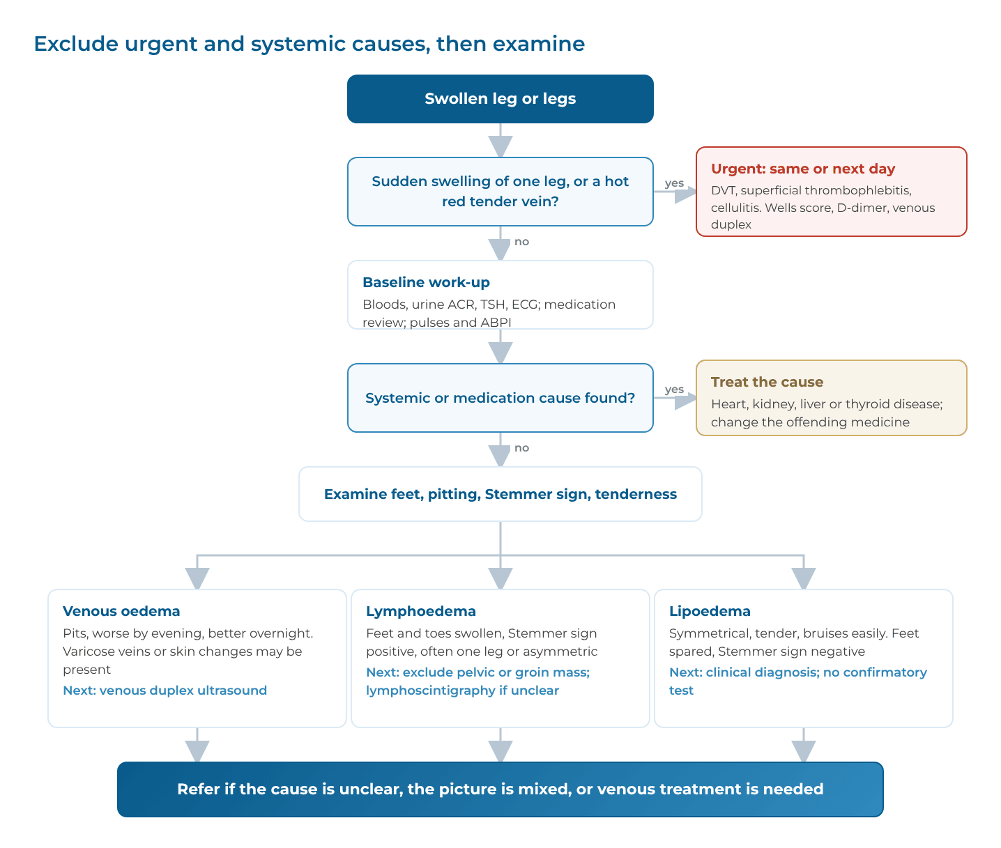
Tests are chosen to exclude systemic and venous causes first; lymphoedema and lipoedema are then largely clinical diagnoses supported by targeted imaging.
Primary-care work-up
| Test | Why |
|---|---|
| FBC, UEC/eGFR, LFTs, albumin | Kidney, liver and protein status |
| Urinalysis and urine albumin:creatinine ratio | Nephrotic-range proteinuria |
| TSH | Hypothyroidism (also more common in lipoedema) |
| HbA1c, lipids | Metabolic risk, especially with obesity or lipoedema |
| ECG; BNP/NT-proBNP and echocardiogram if cardiac features | Heart failure, pulmonary hypertension |
| Wells score ± D-dimer, then urgent venous duplex | Suspected DVT or superficial thrombophlebitis (acute swelling, painful cord-like vein) |
| Venous duplex ultrasound (reflux study) | Chronic venous insufficiency, post-thrombotic change; the most useful single imaging test for chronic leg swelling |
| Medication review | Calcium-channel blockers, gabapentinoids, NSAIDs, steroids, hormones |
| Limb circumference at fixed points, photos | Baseline to track change |
Specialist tests (as indicated)
- Lymphoscintigraphy: the standard functional test of lymphatic drainage; confirms lymphoedema when the clinical picture is uncertain
- Indocyanine green (ICG) lymphography: maps superficial lymphatics, shows dermal backflow; used to plan lymphatic surgery
- MR lymphangiography: detailed anatomy in complex or primary lymphoedema
- CT or MR venography, intravascular ultrasound (IVUS): iliac vein compression or obstruction (e.g. May-Thurner)
- CT or MRI of abdomen and pelvis: new unilateral lymphoedema in an adult, to exclude a mass or recurrent cancer
- Ultrasound of the subcutaneous tissue: fat thickness and echotexture; can support (not prove) lipoedema
- Bioimpedance spectroscopy: detects early extracellular fluid build-up, mainly used in cancer-related lymphoedema surveillance
There is no blood test or scan that confirms lipoedema; the diagnosis rests on history, examination and excluding other causes.
Red flags: refer urgently
- Acute painful swelling of one leg (possible DVT), or a hot, red, tender cord-like vein (superficial thrombophlebitis) – duplex the same or next day
- Hot, red, spreading swelling with fever (cellulitis or sepsis)
- New swelling with breathlessness, chest pain or rapid weight gain
- New unilateral swelling in an adult with weight loss, a past cancer or a palpable pelvic or groin mass
- Rapidly progressive swelling, ulceration, or a cold, pale foot
Treatment options
Compression, movement and skin care help all three conditions, but the underlying cause decides everything else. Check the arterial circulation (pulses ± ABPI) before prescribing firm compression to anyone.
Oedema
Treat the cause
- Optimise heart failure, kidney, liver and thyroid disease; diuretics are appropriate only for systemic fluid overload – they do not fix venous, lymphatic or drug-induced swelling and can cause dehydration and kidney injury
- Reduce or swap offending medicines (e.g. amlodipine to an alternative antihypertensive; review gabapentinoids)
- Anticoagulation for DVT; duplex-guided treatment for superficial thrombophlebitis (compression, anti-inflammatories, and anticoagulation for longer or proximal segments)
Conservative (venous and dependent oedema)
- Graduated compression stockings – usually 20-30 mmHg below-knee for symptomatic venous disease, and 30-40 mmHg for skin changes, healed ulcers or post-thrombotic syndrome – put on first thing in the morning. Correct fit is essential: ankle, calf and (for thigh-length) thigh circumferences and leg length are measured, ideally early in the day when swelling is least. The Perth Vascular Clinic Stockings service provides measurement, fitting, and advice on application aids and garment replacement (usually every 4-6 months)
- Leg elevation above heart level, calf-muscle exercise, walking, avoiding prolonged sitting
- Weight reduction; skin moisturising; prompt treatment of eczema
Endovenous and surgical (for venous causes)
- Endovenous thermal ablation (radiofrequency or laser) of incompetent saphenous veins – minimally invasive, local anaesthetic, now first-line over open stripping
- Non-thermal options: cyanoacrylate glue closure, ultrasound-guided foam sclerotherapy
- Phlebectomy for residual varicose veins
- Iliac vein stenting for symptomatic iliac obstruction (May-Thurner or post-thrombotic), selected on venography and IVUS
- Open venous surgery – high ligation and stripping of the great or small saphenous vein (for example very large, tortuous or near-surface veins unsuitable for ablation, or recurrent varicose veins), open ligation of incompetent perforator veins, and, in specialist centres, deep venous reconstruction (valve repair, endophlebectomy or venous bypass) for severe post-thrombotic disease
Treating venous reflux reduces swelling, improves symptoms and speeds ulcer healing; it also lowers the risk of venous ulcers recurring.
Lymphoedema
There is no cure, but early, consistent treatment controls it well.
Complete (complex) decongestive therapy – CDT
Delivered by an accredited lymphoedema practitioner:
- Intensive phase: multilayer short-stretch bandaging, manual lymphatic drainage, exercise and skin care over several weeks to reduce volume
- Maintenance phase: made-to-measure flat-knit compression garments, self-massage, exercise, skin care; garments replaced every 6 months or so
Adjuncts
- Intermittent pneumatic compression (selected patients, home use)
- Weight management – obesity worsens lymphoedema and can cause it
- Meticulous skin and nail care; treat tinea between the toes
- Cellulitis: treat promptly; for recurrent episodes, long-term prophylactic penicillin per Therapeutic Guidelines
Surgery (specialist lymphatic centres)
- Lymphaticovenular anastomosis (LVA): microsurgical bypass of lymphatics into small veins; best in early, fluid-predominant disease
- Vascularised lymph node transfer (VLNT): transplant of healthy nodes to the affected limb
- Suction-assisted protein lipectomy (lymphoedema liposuction) – for late, non-pitting, fat-dominant swelling; requires lifelong compression afterwards
- Excisional (debulking) operations – reserved for severe stage III disease
Lipoedema
Treatment aims to relieve pain, maintain mobility and slow progression; it does not eliminate the underlying disease. The 2026 consensus recommends individualised, multidisciplinary care.
Conservative – first line for everyone
- Compression: flat-knit garments reduce pain, heaviness and bruising for many women; fit and comfort matter more than maximum pressure
- Exercise: low-impact activity, especially water-based exercise (aqua aerobics, swimming), walking, cycling, yoga
- Nutrition and weight optimisation: prevents coexisting obesity from worsening the legs; anti-inflammatory Mediterranean-style or lower-carbohydrate approaches are commonly used, though lipoedema fat itself is diet-resistant. Track waist-to-height ratio rather than BMI alone
- CDT / manual lymphatic drainage: helpful in some patients, particularly with heaviness or lymphatic overload; not every component is needed
- Psychological support: body image, anxiety, depression and coping; connection with patient groups such as Lipoedema Australia
Medicines
No medicine has proven effectiveness against lipoedema itself. GLP-1-based weight-loss medicines and bariatric surgery treat coexisting obesity – often worthwhile – but the lipoedema component and its pain typically persist.
Surgery
- Lymph-sparing liposuction (tumescent, water-jet or power-assisted techniques, usually in stages) removes the abnormal fat and can improve pain, bruising, mobility and quality of life in selected patients
- Evidence comes mainly from uncontrolled cohort studies; randomised trials and long-term data are still lacking
- Where obesity and metabolic disease coexist, these should generally be treated first
- Surgery should be done by teams experienced in lipoedema, as part of ongoing conservative care – not as a cosmetic procedure
- In Australia, Medicare and private-fund coverage for lipoedema surgery is limited and criteria-specific; patients should confirm eligibility and costs beforehand
- Excision of overhanging skin folds may be needed after major volume reduction
Outcomes and what happens if untreated
All three are chronic; outcomes depend on the cause, how early it is recognised, and how consistently treatment is kept up.
| Condition | With treatment | If untreated |
|---|---|---|
| Oedema | Usually resolves or is well controlled once the cause is treated. Venous ablation gives lasting symptom relief in most patients; drug-induced oedema settles after the medicine is changed | Persistent venous hypertension leads to skin damage – pigmentation, eczema, lipodermatosclerosis – and eventually venous ulcers. Long-standing oedema can overload the lymphatics (phlebolymphoedema). Untreated DVT risks pulmonary embolism and post-thrombotic syndrome |
| Lymphoedema | Early (stage I) disease can be reduced substantially and held stable with garments and self-care. Lymphatic microsurgery can reduce or eliminate garment needs in selected early cases | Progressive: fluid gives way to fat and fibrosis, the limb enlarges and hardens, and changes become irreversible. Recurrent cellulitis (each episode further damages lymphatics), skin breakdown, lymph leakage, reduced mobility. Rarely, after many years, a lymphangiosarcoma (Stewart-Treves syndrome) |
| Lipoedema | Compression, exercise and weight optimisation reduce pain and heaviness and protect mobility. After liposuction, published cohorts report sustained improvement in pain, bruising and quality of life for several years in most patients | Course varies and progression is not inevitable. Worsening is linked to weight gain and hormonal events. Advanced disease brings lobular deformity, gait change, knee and hip osteoarthritis, secondary lymphoedema (lipo-lymphoedema) and significant psychological distress |
Complications and side effects of treatment
Most treatments are low risk; the main hazards are compression in people with poor arterial circulation, misuse of diuretics, and the general risks of surgery.
| Treatment | Possible side effects and complications |
|---|---|
| Compression stockings and bandaging | Discomfort, heat, skin irritation and allergy, difficulty applying; pressure injury or skin necrosis if arterial supply is poor or the garment is ill-fitting – contraindicated in significant peripheral arterial disease |
| Diuretics | Dehydration, low sodium or potassium, gout, acute kidney injury, falls – and no benefit for venous, lymphatic, lipoedema or drug-induced swelling |
| Endovenous ablation (RFA / laser) | Bruising, tenderness along the vein, temporary numbness (saphenous or sural nerve), superficial phlebitis, skin pigmentation; uncommon DVT or heat-induced thrombus extension (checked with early duplex) |
| Open venous surgery (ligation and stripping, perforator ligation) | Bruising, haematoma, wound infection, groin lymph leak or lymphocele, numbness from saphenous or sural nerve injury, DVT; recurrence from new vein growth (neovascularisation); anaesthetic risks and a longer recovery than endovenous treatment |
| Cyanoacrylate glue | Phlebitis-like inflammatory reaction along the vein; rare hypersensitivity |
| Foam sclerotherapy | Pigmentation, matting (fine new vessels), local thrombus; rare visual disturbance or migraine-like symptoms; rare DVT |
| Iliac vein stenting | Groin haematoma, back pain, stent thrombosis or narrowing, need for repeat procedures; anticoagulation-related bleeding |
| CDT and manual lymphatic drainage | Time burden, bandage discomfort, skin irritation; should be avoided during active cellulitis, untreated DVT or decompensated heart failure |
| Pneumatic compression | Discomfort; may shift fluid to the genital area if the pelvis is not treated; contraindicated in active DVT, infection and heart failure |
| Lymphatic microsurgery (LVA, VLNT) | Wound infection, seroma, failure of the bypass or graft; with node transfer, risk of new lymphoedema at the donor site |
| Liposuction for lipoedema or lymphoedema | Bruising, swelling, pain, seroma, infection, contour irregularity, numbness, loose skin; fluid and blood loss with large-volume sessions; DVT and pulmonary embolism; rarely fat embolism or local-anaesthetic toxicity. Lymphatic injury is minimised by lymph-sparing technique. Lymphoedema patients need lifelong compression afterwards |
| Weight-loss medicines and bariatric surgery | Nausea, gastrointestinal upset, gallstones, nutritional deficiencies; lipoedema symptoms often persist |
When to refer, and how we can help
Suggested referral to a vascular surgeon
- Chronic leg swelling where the cause is unclear after initial work-up
- Varicose veins, venous skin changes or ulceration; recurrent superficial thrombophlebitis
- Unilateral swelling, particularly of the left leg, suspicious of iliac vein compression or post-thrombotic obstruction
- Suspected lymphoedema or lipoedema needing confirmation, staging or a coordinated treatment plan
- Mixed pictures – e.g. lipoedema with venous disease, or phlebolymphoedema
- Swelling with reduced pulses or an abnormal ABPI before compression is started
Refer acute DVT, superficial thrombophlebitis near the groin or knee, cellulitis with sepsis, cardiac failure and suspected malignancy along urgent pathways first. Our GP referral guidelines cover other vascular conditions.
At Perth Vascular Clinic
We offer a single assessment that separates venous, lymphatic and fat-related causes of leg swelling:
- Consultation and examination, including ABPI where appropriate (see our ABI clinic)
- Diagnostic vascular ultrasound through the Perth Vascular Clinic Ultrasound service or partner service providers – venous reflux and obstruction studies, urgent duplex for suspected DVT or superficial thrombophlebitis, and assessment of the subcutaneous tissue
- Further imaging when needed – CT/MR venography, IVUS, lymphoscintigraphy or ICG lymphography through partner service providers
- Treatment of venous causes – endovenous ablation, cyanoacrylate closure, sclerotherapy, phlebectomy, open venous surgery and iliac vein stenting
- Perth Vascular Clinic Stockings service – measurement and fitting of graduated compression stockings, with advice on wear and replacement
- Coordination with accredited lymphoedema practitioners, dietitians, psychologists and, where indicated, specialist lymphatic or lipoedema surgical teams
Key messages for patients
- Swollen legs have many causes – not all swelling is “fluid”, and not all large legs are due to weight
- Oedema usually has a treatable cause; it often improves overnight and pits when pressed
- Lymphoedema is lifelong but controllable – early treatment and good skin care prevent progression and infection
- Lipoedema is a real medical condition: painful, symmetrical fat that spares the feet and resists dieting. It is not your fault
- Compression, regular low-impact exercise and a healthy weight help all three
- See a doctor urgently if one leg becomes suddenly swollen and painful, or red and hot with fever
Arranging a consultation
Perth Vascular Clinic offers a single assessment for swollen legs that separates venous, lymphatic and fat-related causes, with vascular ultrasound, the full range of endovenous and surgical treatments for venous disease, a compression stockings fitting service, and coordination with lymphoedema and lipoedema specialists.
Dr Marek Garbowski, FRACS (Vasc). Suite 218, St John of God Medical Clinic, 25 McCourt Street, Subiaco WA 6008. Tel 08 6116 4955
Request an AppointmentCall 08 6116 4955References
- Kruppa P, Crescenzi R, Faerber G, et al. Lipedema World Alliance Delphi consensus-based position paper on the definition and management of lipedema. Nat Commun. 2026;17:427. doi:10.1038/s41467-025-68232-z
- International Society of Lymphology. The diagnosis and treatment of peripheral lymphedema: 2023 consensus document of the International Society of Lymphology. Lymphology. 2023;56(4):133-151. doi:10.2458/lymph.6372
- Faerber G, Cornely M, Daubert C, et al. S2k guideline lipedema. J Dtsch Dermatol Ges. 2024. (German Society of Phlebology and Lymphology)
- Amato ACM, Peclat APRM, Kikuchi R, et al. Brazilian consensus statement on lipedema using the Delphi methodology. J Vasc Bras. 2025;24:e20230183.
- Vazirnia A, Smart DR, Mohseni Y, Amron DM. Lipedema diagnosis, clinical manifestations, and therapeutics: a systematic review. Int J Dermatol. 2026. doi:10.1111/ijd.70227
- De Maeseneer MG, Kakkos SK, Aherne T, et al. European Society for Vascular Surgery (ESVS) 2022 clinical practice guidelines on the management of chronic venous disease of the lower limbs. Eur J Vasc Endovasc Surg. 2022;63(2):184-267.
- Gohel MS, Heatley F, Liu X, et al. A randomized trial of early endovenous ablation in venous ulceration (EVRA). N Engl J Med. 2018;378:2105-2114.
- National Institute for Health and Care Excellence. Liposuction for chronic lipoedema. Interventional procedures guidance IPG721. 2022.
- Therapeutic Guidelines (eTG). Antibiotic: cellulitis and erysipelas, including prophylaxis of recurrent cellulitis. Therapeutic Guidelines Ltd, current edition.
General information only. This article provides general information and does not replace individual medical advice. Please discuss your own situation with your GP or specialist. All endovascular and surgical procedures carry risks. Before proceeding, you may wish to seek a second opinion from an appropriately qualified health practitioner.

Listen to the Latest Podcast
Listen to the latest podcast featuring Dr Marek Garbowski and Medical Forum, WA’s independent monthly publication for health professionals.
Get in Touch

About the Author: Dr Marek Garbowski
Dr Marek Garbowski, MBBS, FRACS (Vasc), is a Vascular and Endovascular Surgeon and the founder of Perth Vascular Clinic, which he established in Subiaco in 2006.
He completed his MBBS in 1994 and trained across Australia and New Zealand before being awarded Fellowship of the Royal Australasian College of Surgeons in Vascular Surgery in 2005. He performs both open vascular surgery and minimally invasive endovascular procedures, with particular interests in carotid stenting, complex aortic aneurysm repair, endovenous treatment of varicose veins and the management of leg and foot ulcers.
Dr Garbowski consults in Subiaco and Joondalup, heads the Vascular and Endovascular Surgery Department at Joondalup Health Campus, and teaches at UWA and Notre Dame.

Millions of Women suffer from Pelvic Pain
In some cases, this pain can be caused by Pelvic Venous Disorders (also known as Pelvic Congestion Syndrome).
If you're in pain and can't seem to find any answers, read our guide and find out more.