
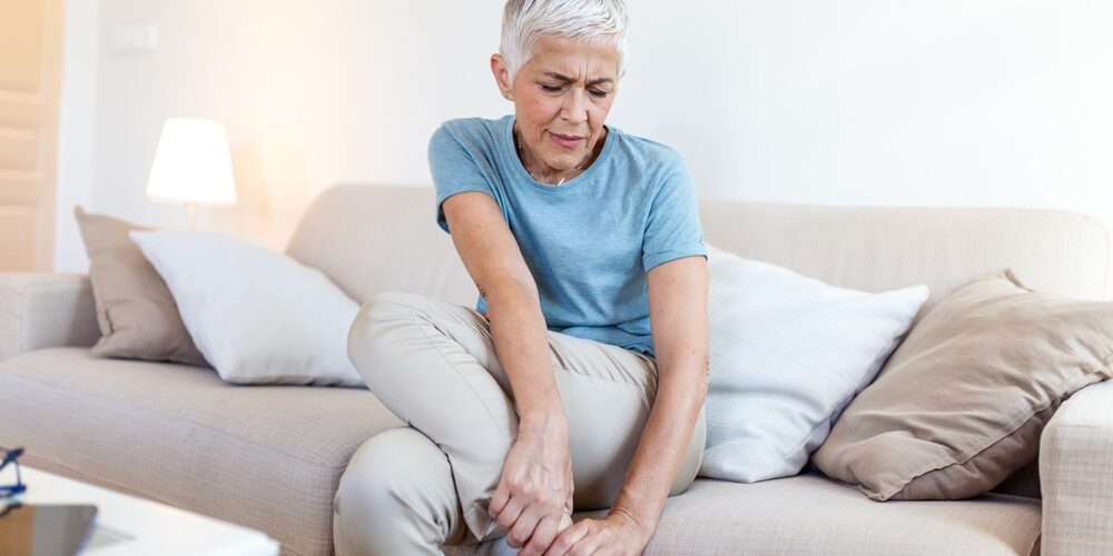
Aching legs are one of the most common reasons people see their GP. Most of the time the cause is harmless, such as prolonged standing, unaccustomed exercise or simple fatigue. For a significant number of people, though, the ache is the first sign of a circulation problem. This can be in the veins, where blood struggles to return to the heart, or in the arteries, where not enough blood reaches the muscles.
Telling these apart matters. Venous disease is mostly a quality-of-life problem that can progress to skin damage and ulceration. Arterial disease is a marker of widespread atherosclerosis and carries a real risk of heart attack and stroke. This article explains how the different causes present, what a vascular assessment involves, which tests are useful, and what treatment options exist.
The short version
- Venous aching builds through the day and eases with walking, elevation or compression.
- Arterial aching (claudication) is a cramping pain on walking that settles within minutes of standing still.
- Pain in the foot at rest, ulcers or gangrene need urgent vascular referral.
- A standing venous duplex and an ankle-brachial index (ABI) answer most questions.
- Peripheral arterial disease is a marker of heart attack and stroke risk, so risk-factor control comes first.
How common is it?
Chronic venous disease is extremely common. Large population studies from Edinburgh and Bonn found that most adults have at least some visible veins such as spider veins. Varicose veins affect roughly one in four to one in three adults, and symptoms of heaviness and aching are reported by a large share of the population. Prevalence rises with age, female sex, pregnancy, obesity, family history and occupations that involve prolonged standing.
Peripheral arterial disease (PAD) affects about one in five people over 60–65, although only around a third of them have typical symptoms. Smoking and diabetes are the strongest risk factors, followed by hypertension, high cholesterol, chronic kidney disease and age.
What causes aching legs?
The causes fall into vascular and non-vascular groups. The pattern of symptoms, especially their timing and what makes them better or worse, is usually the most useful clue.
| Cause | Typical pattern | Relieved by | Key clues |
|---|---|---|---|
| Chronic venous insufficiency / varicose veins | Heaviness, aching, throbbing, itching; worse at end of day and in heat | Elevation, walking, compression | Visible veins, ankle swelling, skin changes |
| Pelvic venous disorders | Leg heaviness plus pelvic ache; vulval, perineal or posterior thigh varices | Lying down | Symptoms worse after standing, around menstruation or after intercourse |
| Post-thrombotic syndrome | Heaviness, swelling, “bursting” pain on walking | Elevation | Prior DVT, persistent swelling |
| Peripheral arterial disease (intermittent claudication) | Cramping calf, thigh or buttock pain on walking, at a reproducible distance | Standing still for 1–5 minutes | Weak or absent pulses, smoking or diabetes |
| Critical limb-threatening ischaemia | Pain in the foot at rest, worse at night | Hanging the leg over the bed edge | Ulcers, gangrene, very low ankle pressures |
| Neurogenic claudication (spinal stenosis) | Aching, numbness or weakness in the legs on standing or walking | Sitting or bending forward (e.g. over a shopping trolley) | Normal pulses, back pain |
| Popliteal artery entrapment / chronic exertional compartment syndrome | Calf pain in young, active people during exercise | Rest | Athletes, normal resting circulation |
| Musculoskeletal (osteoarthritis, tendinopathy, Baker’s cyst) | Joint-related, variable with movement | Variable | Localised tenderness, joint signs |
| Lipoedema | Aching, tenderness, easy bruising of symmetrical fatty legs, sparing the feet | Poorly relieved | Women, onset at puberty or pregnancy |
| Restless legs syndrome | Uncomfortable urge to move, worse in the evening and at rest | Movement | Often linked to low iron stores |
| Medication or metabolic causes | Diffuse muscle aches | Stopping or correcting the cause | Statins, hypothyroidism, vitamin D deficiency |
| Deep vein thrombosis (acute) | New one-sided pain and swelling | Not relieved | Urgent assessment needed |
Presentation: the two main vascular patterns
Venous aching tends to build through the day. Patients describe heavy, tired or restless legs that are worst in the evening, after long periods of standing or sitting, and in hot weather. Symptoms often ease with walking, elevation or compression stockings. Night cramps, itching and ankle swelling are common. In women, symptoms often fluctuate with the menstrual cycle and worsen with each pregnancy.
Leg heaviness together with pelvic pain or varicose veins in unusual places (the vulva, the back of the thigh) suggests an underlying pelvic venous disorder. This group is often missed and needs specific assessment. Read more about pelvic venous disease
Arterial aching (intermittent claudication) is different. It is a cramping pain that comes on with exercise, usually in the calf, occurs at a fairly predictable walking distance and goes away within a few minutes of standing still. The location of the pain reflects the level of the blockage: buttock and thigh pain suggests disease in the aorta or iliac arteries, while calf pain suggests narrowing of the femoral or popliteal arteries. Our guide to peripheral arterial disease explains the condition, its tests and its treatment in more detail.
Pain at rest in the foot, particularly at night and relieved by hanging the leg out of bed, is a warning sign of critical ischaemia and needs urgent referral.
Examination
A vascular examination should be done with the patient both standing and lying.
With the patient standing, the legs are inspected for spider veins, reticular and varicose veins, ankle swelling, and skin changes such as brown pigmentation, eczema, hardening of the skin (lipodermatosclerosis), white scarring (atrophie blanche) and ulceration. The groin, vulva and back of the thigh should be examined for atypical varices that point to a pelvic source. Findings are recorded using the internationally recognised CEAP classification (Clinical, Etiological, Anatomical, Pathophysiological), which grades venous disease from C0 (no visible signs) to C6 (active ulcer).
With the patient lying, the femoral, popliteal, posterior tibial and dorsalis pedis pulses are palpated and the abdomen is examined for an aneurysm. The feet are checked for colour, temperature, capillary refill, hair loss, shiny skin, nail changes, and any ulcers or gangrene between the toes and on the heels. Buerger’s test (pallor when the leg is raised, followed by dusky red discolouration when it is lowered) indicates significant arterial disease.
A brief back, hip and knee examination, along with checks of reflexes and sensation, helps identify spinal, joint and neuropathic causes.
Investigations
Ankle-brachial index (ABI). This simple, non-invasive test compares blood pressure at the ankle with blood pressure in the arm. A value below 0.90 confirms PAD. In people with diabetes or kidney disease, calcified arteries can falsely raise the ABI, so a toe-brachial index is more reliable. A normal resting ABI in someone with typical symptoms should be followed by an exercise ABI.
Venous duplex ultrasound. This is the key test for venous disease. It maps which veins are leaking (reflux), how severe the leak is, and whether there is past or present clot. It must be done standing, by an experienced vascular sonographer, and is essential before planning any venous treatment. Where a pelvic source is suspected, transvaginal or transabdominal pelvic venous duplex can show pelvic varices and reflux in the ovarian and internal iliac veins.
Arterial duplex ultrasound. This shows the location and severity of arterial narrowings and blockages without radiation or contrast.
CT or MR angiography is reserved for patients in whom intervention is being planned. Catheter angiography is now mainly used at the time of treatment.
Blood tests help with the overall assessment and with excluding other causes. They include full blood count, kidney function, HbA1c, lipid profile, and, where relevant, TSH, CK (particularly on statins), vitamin D and ferritin (for restless legs).
MRI of the lumbar spine is appropriate when the history suggests spinal stenosis and the circulation tests are normal.
Assessment pathway
Natural history: what happens without treatment?
Venous disease tends to progress slowly. Long-term follow-up from the Bonn Vein Study found that roughly one in five people with varicose veins progressed to more advanced disease, such as swelling or skin changes, over about six to seven years. Once skin changes develop, the risk of ulceration increases. Venous ulcers heal slowly and commonly recur. Superficial thrombophlebitis and, less often, bleeding from a varix are other complications. Venous disease does not shorten life, but it can significantly affect comfort, work and quality of life.
Intermittent claudication is, in the legs themselves, more benign than many patients fear. Most people remain stable or improve over five years, particularly if they stop smoking and exercise. Only a minority deteriorate, and major amputation is uncommon, in the order of 1–2% over five years. However, PAD is a strong marker of disease elsewhere in the body, and a substantial proportion of patients have a heart attack or stroke within five years. This is why aggressive cardiovascular risk reduction is the most important part of treatment.
Critical limb-threatening ischaemia, by contrast, has a high risk of amputation without revascularisation.
Management
Venous disease
Conservative measures include regular walking, calf-muscle exercise, weight management, elevating the legs when resting, and graduated compression stockings (usually class 1–2, 15–30 mmHg). Compression relieves symptoms but does not cure the underlying problem, and many patients find it hard to wear stockings in the Perth climate.
Endovenous thermal ablation (endovenous laser ablation or radiofrequency ablation) is the first-line treatment for leaking saphenous veins in international guidelines, including the 2022 European Society for Vascular Surgery (ESVS) guidelines. It is performed under local anaesthetic through a needle puncture, with ultrasound guidance, and most patients return to normal activities within a day or two.
Cyanoacrylate closure (medical glue) seals the vein without heat and without the need for multiple local anaesthetic injections. Compression afterwards is often unnecessary.
Ultrasound-guided foam sclerotherapy involves injecting a sclerosant foam into the vein under ultrasound guidance. It is well suited to recurrent varicose veins, tortuous tributaries and smaller veins.
Ambulatory phlebectomy removes surface varicose veins through tiny incisions and is often combined with ablation.
Open surgery (high ligation and stripping) is now used much less often but still has a place in selected cases.
For patients with venous ulcers, the EVRA trial showed that treating the superficial venous reflux early, rather than delaying until the ulcer has healed, speeds healing and reduces recurrence.
Pelvic venous disorders are treated by embolisation of the leaking ovarian and internal iliac veins, a minimally invasive procedure through a small puncture in the neck or groin. Where there is compression of the iliac vein, venous stenting may be considered.
Peripheral arterial disease
Best medical therapy is essential for every patient. It includes stopping smoking, a high-intensity statin, an antiplatelet agent (aspirin or clopidogrel), and good control of blood pressure and diabetes. In selected patients, the combination of low-dose rivaroxaban (2.5 mg twice daily) with aspirin further reduces cardiovascular and limb events, based on the COMPASS and VOYAGER-PAD trials.
Supervised exercise therapy is the most effective single treatment for claudication. Regular walking to the point of moderate discomfort, three times a week for at least three months, typically improves walking distance substantially. Cilostazol may help some patients but is not suitable for people with heart failure.
Endovascular treatment (balloon angioplasty, with or without stenting or drug-coated devices) is performed through a needle puncture. It is considered when symptoms remain disabling despite medical and exercise therapy, and it is required for critical ischaemia.
Open surgical treatment, such as endarterectomy or bypass using the patient’s own vein or a synthetic graft, is used for longer or more complex blockages. It remains the standard for many patients with extensive disease, in line with the Global Vascular Guidelines for chronic limb-threatening ischaemia.
Complications and side effects of treatment
Compression can cause skin irritation, discomfort and, in people with unrecognised arterial disease, pressure damage. This is why the ABI should be checked before prescribing stockings.
Thermal ablation and glue commonly cause bruising, tenderness and tightness along the treated vein. Less common problems include superficial thrombophlebitis, skin pigmentation, numbness from injury to a nearby nerve (particularly with below-knee treatment), and clot extending into the deep veins (endothermal heat-induced thrombosis, or EHIT). DVT and pulmonary embolism are rare, generally occurring in fewer than 1–2% of cases. Glue can occasionally cause a local inflammatory or allergic reaction.
Foam sclerotherapy can cause pigmentation, matting (fine new spider veins), thrombophlebitis and, rarely, temporary visual disturbance, migraine-like symptoms or DVT.
Pelvic vein embolisation carries small risks of coil migration, post-procedure pelvic pain, and puncture-site bruising.
Angioplasty and stenting may be complicated by puncture-site bleeding or false aneurysm, contrast reactions or kidney injury, arterial dissection, or later re-narrowing (restenosis), which may need repeat treatment.
Bypass surgery carries risks of wound infection, leg swelling, graft blockage, and the cardiac and general risks of major surgery.
When should a GP refer?
| Referral | When |
|---|---|
| Urgent | Suspected critical limb-threatening ischaemia (rest pain, ulceration or gangrene); acute limb ischaemia (sudden pain, pallor and loss of pulses, which is an emergency); suspected DVT. |
| Routine | Lifestyle-limiting claudication; symptomatic varicose veins; skin changes of chronic venous disease; venous ulcers (which should be referred early, in line with the EVRA findings); recurrent varicose veins; bleeding varicose veins; suspected pelvic venous disorders; and any patient in whom the cause of leg symptoms remains unclear after initial assessment. |
Arranging a consultation
Perth Vascular Clinic offers comprehensive assessment of aching legs, including on-site vascular duplex ultrasound and ABI testing, and the full range of treatment options, from compression and minimally invasive venous procedures to endovascular and open arterial surgery.
Suite 218, St John of God Medical Clinic, 25 McCourt Street, Subiaco WA 6008. Tel 08 6116 4955
Request an AppointmentCall 08 6116 4955This article provides general information only and does not replace individual medical advice. Please consult your GP or vascular specialist about your own circumstances.

Listen to the Latest Podcast
Listen to the latest podcast featuring Dr Marek Garbowski and Medical Forum, WA’s independent monthly publication for health professionals.
Get in Touch

About the Author: Dr Marek Garbowski
Dr Marek Garbowski, MBBS, FRACS (Vasc), is a Vascular and Endovascular Surgeon and the founder of Perth Vascular Clinic, which he established in Subiaco in 2006.
He completed his MBBS in 1994 and trained across Australia and New Zealand before being awarded Fellowship of the Royal Australasian College of Surgeons in Vascular Surgery in 2005. He performs both open vascular surgery and minimally invasive endovascular procedures, with particular interests in carotid stenting, complex aortic aneurysm repair, endovenous treatment of varicose veins and the management of leg and foot ulcers.
Dr Garbowski consults in Subiaco and Joondalup, heads the Vascular and Endovascular Surgery Department at Joondalup Health Campus, and teaches at UWA and Notre Dame.

Millions of Women suffer from Pelvic Pain
In some cases, this pain can be caused by Pelvic Venous Disorders (also known as Pelvic Congestion Syndrome).
If you're in pain and can't seem to find any answers, read our guide and find out more.