
What peripheral arterial disease is, how it presents, how it is diagnosed and how it is treated, from best medical therapy and exercise through to endovascular and open surgery. Written for patients and referring GPs.
In this article
In brief
- Peripheral arterial disease (PAD) is narrowing or blockage of the arteries to the legs, usually caused by atherosclerosis, the same process behind heart attacks and strokes.
- The classic symptom is claudication: cramping pain in the calf, thigh or buttock on walking that settles within a few minutes of standing still. Many people have no leg symptoms at all.
- Rest pain in the foot, non-healing ulcers or gangrene signal chronic limb-threatening ischaemia and need urgent specialist assessment. A suddenly cold, painful, pale leg is an emergency.
- The ankle-brachial index (ABI) is the first-line test, and arterial duplex ultrasound maps the disease when treatment is being planned.
- Every patient needs best medical therapy: stopping smoking, a statin, an antiplatelet, and control of blood pressure and diabetes. Supervised exercise is the most effective single treatment for claudication, and angioplasty, stenting or bypass surgery are added when symptoms remain disabling or the leg is threatened.
What is peripheral vascular disease?
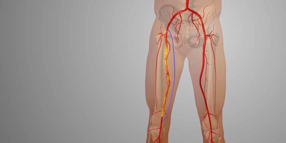
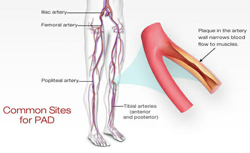
“Peripheral vascular disease” is a broad term for disease of the blood vessels outside the heart and brain. In everyday use, and in this article, it refers to peripheral arterial disease (PAD): narrowing or blockage of the arteries that supply the legs. The main cause is atherosclerosis, a build-up of cholesterol-rich plaque in the artery wall. It is the same process that causes heart attacks and strokes.
PAD matters for two reasons. It can reduce walking ability and, in its severe form, threaten the leg. More importantly, it is a powerful warning sign of atherosclerosis elsewhere in the body. People with PAD have a markedly increased risk of heart attack, stroke and cardiovascular death, and much of the benefit of treatment comes from reducing that risk.
How common is it?
PAD affects more than 200 million people worldwide. Prevalence rises steeply with age, from a few percent in people in their 40s to around one in five in people over 70. Here in Perth, the Health In Men Study found that roughly one in seven men aged 65 and over had an abnormal ankle-brachial index. Many people with PAD have no symptoms or atypical symptoms, so the condition is substantially underdiagnosed.
In Australia, PAD is a major contributor to diabetes-related amputation. Aboriginal and Torres Strait Islander people are disproportionately affected, reflecting higher rates of diabetes and kidney disease.
Causes and risk factors
More than 90% of PAD is due to atherosclerosis. The main risk factors are smoking (the strongest single risk factor), diabetes, high blood pressure, high cholesterol, chronic kidney disease, increasing age, male sex, and a family history of premature cardiovascular disease.
Less common, non-atherosclerotic causes are important to recognise, particularly in younger patients:
- Embolism: clot travelling from the heart (e.g. in atrial fibrillation) or from an aneurysm.
- Popliteal artery aneurysm: a swelling behind the knee that can clot or shed debris. See peripheral aneurysms.
- Popliteal artery entrapment syndrome: compression of the artery by calf muscles in young, athletic people. See arterial and venous compression syndromes.
- Cystic adventitial disease: a rare cyst within the artery wall.
- Thromboangiitis obliterans (Buerger’s disease): inflammation of small arteries in young heavy smokers.
- Vasculitis, fibromuscular dysplasia and radiation arteritis.
How does it present?
PAD ranges from no symptoms at all to limb-threatening disease. Two classification systems are used to grade severity:
| Fontaine stage | Rutherford category | Clinical picture |
|---|---|---|
| I | 0 | No symptoms |
| IIa / IIb | 1–3 | Mild, moderate or severe claudication |
| III | 4 | Ischaemic rest pain |
| IV | 5–6 | Tissue loss: ulceration or gangrene |
Asymptomatic PAD
Many patients have no leg symptoms, often because other conditions (arthritis, heart or lung disease) limit their walking before the legs do. The disease is picked up on examination or testing, but the cardiovascular risk is just as high.
Intermittent claudication
This is the classic symptom: a cramping, aching pain in the calf, thigh or buttock that comes on with walking, at a fairly consistent distance, and settles within a few minutes of standing still. The site of the pain reflects the level of disease:
- Buttock and thigh pain suggests disease in the aorta or iliac arteries, sometimes with erectile dysfunction in men (Leriche syndrome).
- Calf pain suggests narrowing of the femoral or popliteal arteries.
- Foot pain on walking suggests disease below the knee.
Not every leg pain on walking is arterial. Spinal stenosis, venous disease and joint problems can look similar, and our article Aching legs: when should you worry about your veins or arteries? sets out how to tell them apart.
Chronic limb-threatening ischaemia (CLTI)
This is the most severe form. It presents as rest pain in the foot, typically at night and relieved by hanging the leg over the edge of the bed, or as non-healing ulcers or gangrene, often on the toes, heels or between the toes. In people with diabetes, nerve damage can mask pain, so tissue loss may be the first sign. CLTI requires urgent specialist assessment.
Acute limb ischaemia
A sudden blockage, from an embolus or from a clot forming on an existing narrowing, causes the “6 Ps”: pain, pallor, pulselessness, perishing cold, paraesthesia (pins and needles) and paralysis. This is a surgical emergency. Numbness and weakness indicate a threatened limb that may need revascularisation within hours.
A suddenly painful, pale or cold leg is an emergency. If it comes with numbness or weakness, call 000 or go to the nearest emergency department straight away. Do not wait for a routine appointment.
Examination
A vascular examination looks for evidence of reduced blood flow and for the underlying risk factors.
- Pulses: the femoral, popliteal, posterior tibial and dorsalis pedis pulses are felt on both sides. An unusually prominent, easily felt popliteal pulse may indicate an aneurysm.
- Skin and feet: cool, pale or dusky skin, hair loss, shiny thin skin, thickened nails, slow capillary refill, and ulcers or gangrene, particularly between the toes and on the heels.
- Buerger’s test: pallor when the leg is elevated, followed by a dusky red colour (dependent rubor) when it is lowered, indicating severe disease.
- Bruits: a whooshing sound over the femoral arteries or abdomen suggests turbulent flow through a narrowing.
- Abdomen: palpation for an abdominal aortic aneurysm, which often coexists with PAD.
- General assessment: blood pressure in both arms, heart rhythm (atrial fibrillation), and signs of heart, carotid or kidney disease.
Investigations
Ankle-brachial index (ABI)
The ABI compares the blood pressure at the ankle with that in the arm. It is quick, painless and the first-line test for PAD. It is performed on site at our ABI clinic.
| ABI | Interpretation |
|---|---|
| > 1.40 | Non-compressible (calcified) arteries: result unreliable |
| 1.00–1.40 | Normal |
| 0.91–0.99 | Borderline |
| 0.70–0.90 | Mild PAD |
| 0.40–0.69 | Moderate PAD |
| < 0.40 | Severe PAD, often with rest pain or tissue loss |
In people with diabetes or kidney disease, arterial calcification can make the ABI falsely normal or high, so a toe-brachial index or toe pressures are used instead. If the resting ABI is normal but the history is typical of claudication, an exercise ABI (treadmill test) can reveal disease that only shows under exertion. Transcutaneous oxygen measurement helps predict wound healing.
Arterial duplex ultrasound
Duplex ultrasound shows the exact site, length and severity of narrowings and blockages without radiation or contrast. At Perth Vascular Clinic it is the main imaging test used to plan treatment.
CT and MR angiography
CT angiography gives a detailed three-dimensional map of the arteries from the aorta to the feet and is usually performed when intervention is planned. MR angiography is an alternative for some patients. Both require care in patients with reduced kidney function.
Catheter angiography
Digital subtraction angiography is now mainly performed at the time of an endovascular procedure, when the diagnosis and treatment can be carried out together.
Blood tests
These assess risk factors and suitability for treatment: full blood count, kidney function, HbA1c, fasting lipids, and in younger patients or unusual presentations, tests for thrombophilia, inflammatory markers and homocysteine.
Natural history: what happens without treatment?
Claudication
In the legs themselves, claudication usually follows a stable course. Over five years:
- About 70–80% of patients remain stable or improve.
- About 10–20% experience worsening symptoms.
- Only 1–2% progress to major amputation.
The prognosis for the heart and brain is much more serious. Around 15–30% of people with claudication die within five years, mostly from heart attack or stroke. Continued smoking and diabetes greatly increase the risk of deterioration in both the legs and the heart.
Chronic limb-threatening ischaemia
CLTI carries a poor outlook without treatment. Within one year of diagnosis, roughly a quarter to a third of patients who are not revascularised will need a major amputation, and around a quarter will die. Timely revascularisation substantially improves limb salvage.
Acute limb ischaemia
Without prompt treatment, acute limb ischaemia leads to irreversible muscle and nerve damage within hours, with a high risk of amputation and death.
Management
Treatment has two aims: to reduce the risk of heart attack, stroke and death, and to relieve symptoms and save the limb.
1. Best medical therapy (for every patient)
- Stop smoking. This is the single most important measure. It slows disease progression, improves the results of any procedure and reduces amputation and death. Nicotine replacement, varenicline and Quitline support are all effective.
- Statin therapy, usually high-intensity (e.g. atorvastatin 40–80 mg or rosuvastatin 20–40 mg), regardless of cholesterol level. Ezetimibe or PCSK9 inhibitors are added if LDL targets are not met.
- Antiplatelet therapy: aspirin or clopidogrel for all symptomatic patients.
- Low-dose rivaroxaban (2.5 mg twice daily) combined with aspirin further reduces cardiovascular and limb events in selected patients, including after revascularisation, based on the COMPASS and VOYAGER-PAD trials.
- Blood pressure control, preferably including an ACE inhibitor or angiotensin receptor blocker.
- Diabetes control, with SGLT2 inhibitors or GLP-1 receptor agonists offering additional cardiovascular benefit in suitable patients.
- Foot care: daily inspection, well-fitting footwear, regular podiatry, and prompt attention to any wound, particularly in people with diabetes.
2. Exercise therapy (for claudication)
Supervised exercise therapy is the most effective single treatment for claudication. A typical program involves walking to the point of moderate pain, resting, then walking again, for 30–60 minutes at least three times a week, for at least 12 weeks. Most patients substantially increase their pain-free walking distance. Home-based structured walking programs are a reasonable alternative when supervised programs are not available.
Cilostazol can improve walking distance in some patients but must not be used in people with heart failure.
3. Endovascular treatment
Endovascular procedures are performed through a needle puncture, usually in the groin, under local anaesthetic and light sedation, often as a day-case or overnight stay. They include:
- Balloon angioplasty: a balloon is inflated inside the narrowed artery to stretch it open.
- Stenting: a metal mesh tube holds the artery open, particularly useful in the iliac arteries and for longer blockages.
- Drug-coated balloons and drug-eluting stents: these release a medication that reduces re-narrowing, especially in the femoral and popliteal arteries. See drug-eluting balloon angioplasty and drug-eluting self-expanding stents.
- Atherectomy and lithotripsy: specialised devices that remove or fracture heavily calcified plaque.
Endovascular treatment is considered for claudication that remains disabling despite medical and exercise therapy, and is often the first choice for CLTI, particularly in frail patients. More detail is in advanced endovascular management of peripheral vascular disease, and our angiogram, angioplasty and stent discharge information explains what to expect afterwards.
4. Open surgery
- Endarterectomy: the diseased inner lining of the artery is removed, most often at the common femoral artery in the groin.
- Bypass surgery: a new channel is created around the blockage, ideally using the patient’s own vein (usually the great saphenous vein) or a synthetic graft. Examples include aorto-bifemoral, femoro-popliteal and femoro-distal bypass.
Recent major trials have clarified the choice between surgery and endovascular treatment for CLTI. In BEST-CLI, patients with a good-quality saphenous vein had better outcomes with bypass surgery than with endovascular treatment. In BASIL-2, for disease below the knee, a best endovascular treatment-first strategy was associated with better amputation-free survival than a vein bypass-first approach, largely because fewer patients died. In practice, the decision is individualised, taking into account the pattern of disease, the availability of a suitable vein, the patient’s fitness and their preferences. Many patients benefit from a hybrid procedure combining open and endovascular techniques.
5. Amputation
When the leg cannot be revascularised, or when extensive gangrene or infection makes salvage impossible, amputation may be the best way to relieve pain and restore mobility. Modern rehabilitation and prosthetics allow many patients to regain independence. Our major amputation discharge information covers recovery after surgery.
Outcomes of treatment
Results depend on the location and length of the disease, the quality of the arteries beyond the blockage, and ongoing risk-factor control. As a general guide:
- Iliac artery angioplasty and stenting has excellent long-term results, with around 80–90% of arteries remaining open at five years.
- Femoral and popliteal endovascular treatment has higher rates of re-narrowing, although drug-coated technologies have improved durability. Repeat procedures are sometimes needed.
- Femoro-popliteal vein bypass typically has five-year patency of around 70–80%. Synthetic grafts perform less well, particularly below the knee.
- Limb salvage after revascularisation for CLTI is achieved in the majority of patients when treatment is timely.
Stopping smoking and taking statins and antiplatelet medication consistently are the most important factors in keeping arteries and grafts open.
Complications and side effects of treatment
Medications
- Antiplatelets and rivaroxaban: bleeding, including gastrointestinal bleeding and bruising. The risk is higher with combination therapy.
- Statins: muscle aches in a minority of patients, rarely liver enzyme elevation. Most patients tolerate an alternative statin or dose.
- Cilostazol: headache, diarrhoea, palpitations and dizziness.
Endovascular treatment
- Bruising, bleeding or false aneurysm at the puncture site.
- Contrast allergy and contrast-induced kidney injury.
- Arterial dissection, blockage or embolism to the lower leg, occasionally requiring further intervention.
- Re-narrowing (restenosis) or stent fracture over time, sometimes needing repeat treatment.
Open surgery
- Wound infection and wound breakdown, particularly in the groin.
- Leg swelling after bypass.
- Graft blockage or infection.
- Heart attack, stroke, chest infection and other risks of major surgery, which are higher in people with extensive cardiovascular disease.
When should a GP refer?
| Referral | When |
|---|---|
| Emergency (same day) | Suspected acute limb ischaemia. |
| Urgent | Rest pain, non-healing ulcers or gangrene, and diabetic foot wounds with reduced pulses. |
| Routine | Claudication that limits lifestyle despite medical therapy and exercise, abnormal ABI with diagnostic uncertainty, suspected popliteal aneurysm, and young patients with claudication or atypical features. |
Our GP referral guidelines cover other vascular conditions.
Arranging a consultation
Perth Vascular Clinic offers comprehensive assessment of peripheral arterial disease, including on-site arterial duplex ultrasound and ABI testing, and the full range of treatment options: best medical therapy, exercise programs, and minimally invasive endovascular and open surgical reconstruction.
Suite 218, St John of God Medical Clinic, 25 McCourt Street, Subiaco WA 6008. Tel 08 6116 4955
Request an AppointmentCall 08 6116 4955General information only. This article provides general information and does not replace individual medical advice. Please consult your GP or vascular specialist about your own circumstances. All endovascular and surgical procedures carry risks. Before proceeding, you may wish to seek a second opinion from an appropriately qualified health practitioner.

Listen to the Latest Podcast
Listen to the latest podcast featuring Dr Marek Garbowski and Medical Forum, WA’s independent monthly publication for health professionals.
Get in Touch

About the Author: Dr Marek Garbowski
Dr Marek Garbowski, MBBS, FRACS (Vasc), is a Vascular and Endovascular Surgeon and the founder of Perth Vascular Clinic, which he established in Subiaco in 2006.
He completed his MBBS in 1994 and trained across Australia and New Zealand before being awarded Fellowship of the Royal Australasian College of Surgeons in Vascular Surgery in 2005. He performs both open vascular surgery and minimally invasive endovascular procedures, with particular interests in carotid stenting, complex aortic aneurysm repair, endovenous treatment of varicose veins and the management of leg and foot ulcers.
Dr Garbowski consults in Subiaco and Joondalup, heads the Vascular and Endovascular Surgery Department at Joondalup Health Campus, and teaches at UWA and Notre Dame.

Millions of Women suffer from Pelvic Pain
In some cases, this pain can be caused by Pelvic Venous Disorders (also known as Pelvic Congestion Syndrome).
If you're in pain and can't seem to find any answers, read our guide and find out more.