
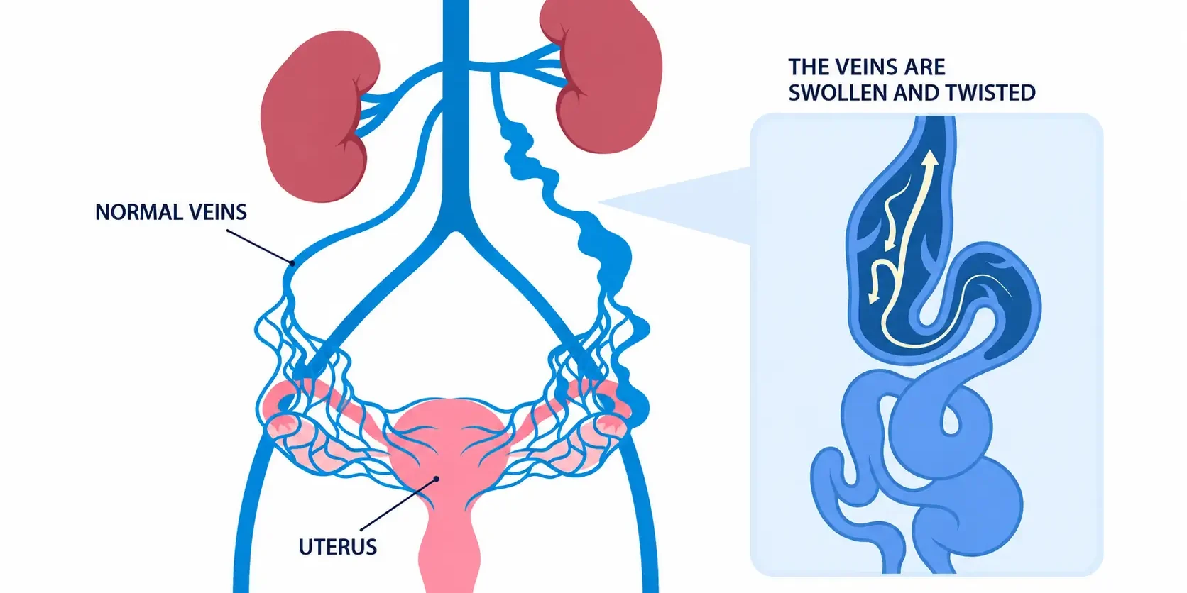
Chronic pelvic pain that has gone on for years and never been given a proper explanation is, more often than people think, coming from the veins. Pelvic venous disease is the second most common cause of chronic pelvic pain, after gynaecological conditions, and it still gets missed for far too long.
The short version
- Pelvic venous disease is the second most common cause of chronic pelvic pain, after gynaecological conditions.
- Symptoms are position and activity dependent, so a scan done at the wrong time of day can come back looking normal.
- Nutcracker and May-Thurner are only worth treating when they are causing symptoms, not because they show up on a scan.
- Ovarian vein embolisation resolves or significantly improves the pain in around 70 to 80 percent of patients.
- A hysterectomy will not fix a venous cause, and in a patient of childbearing age that decision cannot be undone.
Why it gets missed
The biggest gap I see in the work-up of chronic pelvic pain is that a venous cause is dismissed early in the process. It can be investigated quite easily once you think to look for it, with a symptom specific history, transabdominal and transvaginal ultrasound, ovarian vein and testicular vein duplex ultrasound, and CT or MR venogram.
The trouble is that in the early stages of these conditions, the symptoms are position and activity dependent. If a scan is done first thing in the morning, with the patient dehydrated and lying flat and relaxed, it can come back looking normal. I would put the false negative rate at around ten percent. That is not a huge number, but it is not insignificant either, because these are exactly the patients who end up suffering for many years before anyone lands on the right answer. Patients need to be well hydrated, and where possible physically active before the scan, for the result to mean anything.
What it actually feels like
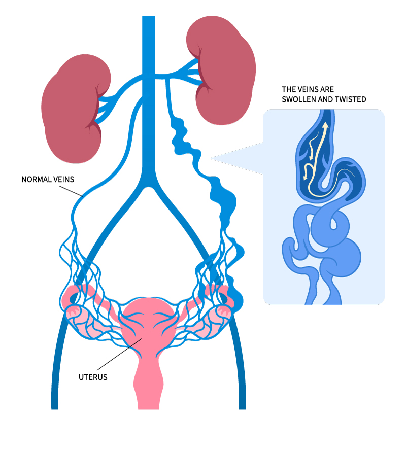
The pain typically builds up gradually over time. It gets worse with physical activity and prolonged standing, and it is relieved, often significantly, by lying flat with the legs elevated. It is not always confined to the lower pelvis either, patients often describe lower back and flank pain as well. Pain during or after intercourse is common, and so is abdominal bloating. None of this happens overnight. What I hear again and again is a pattern of gradual deterioration, something that was manageable becoming less and less so over months or years, to the point where it starts limiting exercise, work and intimacy.
If that pattern sounds familiar, our short pelvic venous disorders quiz is a reasonable place to start, and you can read more about the symptoms of pelvic venous disorders on our website.
Who develops it
Any woman can develop this, but women in their thirties and forties who have had three or more pregnancies are at higher risk. It is not only a condition of women who have had children though. Around ten percent of women are born without competent valves in their ovarian veins, so ovarian vein reflux, and the venous hypertension and pelvic congestion that follow, can affect younger women too, including in their teens and twenties. These younger patients are often the ones who wait longest for a diagnosis, because a venous cause of chronic pelvic pain in a young woman without children is frequently not considered at all. There is also a small number of men who develop pelvic venous discomfort, usually from testicular vein incompetence causing a varicocele, or from May-Thurner syndrome.
How I actually make the diagnosis
I take a detailed, symptom specific history first. That history alone will usually tell me whether the pain is likely to be venous or not. I want other causes properly excluded, particularly gynaecological ones, so a gynaecology review matters. Not every pain is venous. From there I go early to cross sectional imaging, CT venogram or MR venogram, along with a high quality transvaginal ultrasound looking for point tenderness and a dilated network of veins around the ovary.
Where there is genuine doubt about whether a compression seen on imaging is actually causing the patient’s symptoms, I go further, to a diagnostic venogram with intravascular ultrasound, and measurement of the pressure difference between the left renal vein and the inferior vena cava. That combination, imaging plus pressures plus the pattern of collateral veins filling on venogram, is what tells me whether a compression is truly significant, rather than just something that happens to be visible on a scan.
Phenomenon or syndrome
Nutcracker and May-Thurner are anatomical findings. Nutcracker describes compression of the left renal vein just before it joins the inferior vena cava. May-Thurner describes compression of the left common iliac vein where it is squeezed between the right common iliac artery and the sacrum. On their own, seen only on a CT or MR venogram with no symptoms attached, these are phenomena. They are of no clinical consequence, and the patient should be reassured and get on with normal life.
They only become a syndrome, and only then become something to treat, when they are producing real symptoms. For May-Thurner that means pelvic pain, left leg swelling, or a left iliofemoral deep vein thrombosis (DVT). For Nutcracker it means left flank pain, blood or protein in the urine, alongside pelvic pain.
| Nutcracker | May-Thurner | |
|---|---|---|
| Vein compressed | Left renal vein, just before it joins the inferior vena cava | Left common iliac vein, between the right common iliac artery and the sacrum |
| Phenomenon | Seen on a CT or MR venogram with no symptoms attached. Of no clinical consequence, and no treatment needed. | |
| Syndrome | Left flank pain, blood or protein in the urine, alongside pelvic pain | Pelvic pain, left leg swelling, or a left iliofemoral DVT |
| Treatment | Open surgery remains the gold standard. Renal vein stenting still lacks the evidence base. | Iliac vein stenting is the definitive treatment, sometimes venoplasty alone first. |
Even the degree of compression on imaging is not the deciding factor on its own. I have seen more than seventy five percent compression of the left common iliac vein on imaging in a patient with no symptoms at all, and that is not clinically significant. Pressure gradients across a Nutcracker compression are used too, generally somewhere above three to six millimetres of mercury depending which literature you read, but that figure alone does not decide anything either. It is the combination, symptoms, imaging, pressure gradient, and the pattern of collateral veins on a diagnostic venogram, that tells an experienced vascular surgeon whether this is something to treat.
Is it dangerous
Chronic pelvic venous pain itself is not a dangerous condition. It has a real impact on quality of life, which is why we treat it, but it does not threaten life or long term health on its own. Where the risk changes is with confirmed May-Thurner syndrome, which does raise the risk of a left leg deep vein thrombosis, particularly around long haul travel or periods of immobility. I make patients aware of that and advise graduated compression stockings, staying hydrated, and staying mobile on long flights. I do not routinely recommend blood thinning medication for this unless there has already been an actual DVT or significant leg symptoms, such as varicose veins or swelling.
When symptoms flareExercise and travel are otherwise safe, even with mild symptoms. My general advice when symptoms flare is to rest, elevate the legs, use heat packs, and take a simple anti-inflammatory if needed.
The POTS connection
Some of my patients with May-Thurner syndrome have also had episodes of fainting or a racing heart, and ended up with a diagnosis of POTS (postural orthostatic tachycardia syndrome) from a cardiologist before anyone considered the pelvic veins. I have seen a number of these patients improve, sometimes markedly, in their POTS-like symptoms after treatment of the iliac compression. My thinking is that redistribution of venous flow through an overloaded network of collateral veins, along with the volume shift that comes with a large incompetent ovarian vein, may be contributing.
I want to be honest about the strength of that evidence though. It would be fair for a sceptic to say the pain improved, the patient felt better generally, and the POTS symptoms improved for reasons that have nothing to do with the veins themselves. That is a reasonable challenge. My answer is that this is a clinical observation based on a number of patients responding this way, and that there is a body of evidence now suggesting POTS and POTS-like symptoms can be physiologically linked to venous incompetence in the pelvis, or to overload from collateral flow in compression syndromes. It needs more research. It should not be dismissed as coincidence either. POTS still deserves its own cardiology assessment and management regardless.
Treating it without surgery
For patients with milder, early symptoms, I start with lifestyle advice and reassurance that nothing is being damaged by waiting, and that treatment remains available whenever they need it. Venoactive medication, a combination of the flavonoids diosmin and hesperidin, has a body of evidence behind it. I prescribe it for three months before reassessing, because there is a lag of around three months before you see a symptom response. If it works, it can be continued. If it does not, it can simply be stopped and other options considered. Like any medication it is not suitable for everyone, so it is something to discuss with your doctor rather than start on your own.
When intervention makes sense
Where symptoms are already significantly limiting someone’s lifestyle, I will not hesitate to discuss intervention early, rather than insisting on months of conservative treatment first. For ovarian vein incompetence, coil embolisation has, in the published literature and in my experience, around a seventy to eighty percent chance of resolving the pain or significantly improving it, with a low risk of recurrence. It is done through the groin under X-ray guidance, with detachable coils placed and oversized so they will not move, and generally causes minimal complications. Around twenty percent of patients get some post-embolisation inflammation for a few days, which is self limiting and settles with anti-inflammatories and heat packs. Most people are back to normal activity within a couple of days, and I allow four to six weeks to judge the full symptom response. As with any procedure there are risks, and I go through these with each patient before we proceed.
For May-Thurner syndrome, iliac vein stenting is the definitive treatment, though I will sometimes try venoplasty alone first to see how much the compression and its collateral network respond, before committing a patient to a stent. Stenting means anticoagulation for six to twelve months, then lifelong low dose aspirin, and ongoing stent surveillance on a six to twelve monthly basis to catch problems like in-stent narrowing or fracture early. The risk of stent related clot is low, in my experience around one percent, but it is a long term commitment, and I discuss that honestly with patients before we proceed.
Nutcracker syndrome remains the most controversial area. Open surgery, left renal vein transposition or left ovarian vein transposition, is still the gold standard, and it is a significant operation with its own risks of restenosis or failure over time, needing strict surveillance. Renal vein stenting is newer and still without the evidence base, particularly randomised trial evidence, to say it is a safe replacement for open surgery, especially in younger patients who will be living with that stent for decades.
You can read more about the full range of treatment options for pelvic venous disorders on our website.
What changes the outcome
The most costly mistake I seeThe single most costly mistake I have seen is a hysterectomy performed for chronic pelvic pain without first excluding a venous cause. A hysterectomy that does not include removal of the ovaries and ligation of the ovarian veins does nothing for ovarian vein reflux, and nothing for iliac or renal vein compression. In a patient of childbearing age, that decision cannot be undone, and if the real cause was venous, the pain remains exactly where it was.
The part of this that is hard to hear is that these conditions, once they are truly symptomatic, do not resolve on their own. They tend to get worse over time. Once other causes have genuinely been excluded and a venous cause confirmed, waiting rarely helps. My approach is to ask the specific questions early, involve gynaecology to rule out other causes, go early to cross sectional imaging, and be willing to offer intervention early to patients whose symptoms are already affecting their lives, rather than making them wait.
Once other causes have genuinely been excluded and a venous cause confirmed, waiting rarely helps.
Where to from here
If you have had pelvic pain for a long time and no one has looked at your veins, the first step is a conversation with your GP. Ask whether a venous cause has been considered, and whether a referral to a vascular surgeon with an interest in pelvic venous conditions is appropriate. Our questions to ask your doctor handout may help with that conversation. GPs can find our GP information on pelvic congestion syndrome and referral guidelines in the For Doctors section. For more general information on persistent pelvic pain, Jean Hailes for Women’s Health and healthdirect are good independent starting points.
Talk to someone who looks at the veins
You can also listen to my conversation with Medical Forum, Is it pelvic venous congestion syndrome?, or request an appointment with Perth Vascular Clinic.
Request an AppointmentTake the QuizThis article is general information only and reflects the clinical opinion and experience of Dr Marek Garbowski. It is not a substitute for individual medical advice, and it does not replace assessment by your own doctor. Any surgical or invasive procedure carries risks, and outcomes vary between individuals. Before proceeding you should seek a second opinion from an appropriately qualified health practitioner. A referral from your GP is required to see a specialist.

Listen to the Latest Podcast
Listen to the latest podcast featuring Dr Marek Garbowski and Medical Forum, WA’s independent monthly publication for health professionals.
Get in Touch

About the Author: Dr Marek Garbowski
Dr Marek Garbowski, MBBS, FRACS (Vasc), is a Vascular and Endovascular Surgeon and the founder of Perth Vascular Clinic, which he established in Subiaco in 2006.
He completed his MBBS in 1994 and trained across Australia and New Zealand before being awarded Fellowship of the Royal Australasian College of Surgeons in Vascular Surgery in 2005. He performs both open vascular surgery and minimally invasive endovascular procedures, with particular interests in carotid stenting, complex aortic aneurysm repair, endovenous treatment of varicose veins and the management of leg and foot ulcers.
Dr Garbowski consults in Subiaco and Joondalup, heads the Vascular and Endovascular Surgery Department at Joondalup Health Campus, and teaches at UWA and Notre Dame.

Millions of Women suffer from Pelvic Pain
In some cases, this pain can be caused by Pelvic Venous Disorders (also known as Pelvic Congestion Syndrome).
If you're in pain and can't seem to find any answers, read our guide and find out more.